
A lattice-gated exciton pair in α-tubulin with femtosecond electronic coherence at physiological temperature
Justin Echternach1*
1Independent Researcher, United States
* Correspondence:
Justin Echternach
justin.echternach@positivespaces.io
Keywords: tubulin, microtubules, phenylalanine, exciton coupling, electronic coherence, open quantum systems, quantum biology
Abstract
Electronic coherence in proteins is widely expected to vanish at physiological temperature: the environment is too warm, too wet and too noisy. We test that expectation on one aromatic pair, phenylalanines F169 and F202 of α-tubulin, whose rings sit 4.76 Å apart in the microtubule lattice, inside an aromatic relay that couples GTP hydrolysis to lattice compaction (Zehr et al., 2026). Combining fragment and supermolecular time-dependent density functional theory with molecular dynamics of the α monomer, of partial lattices and of closed, nucleotide-bound microtubule rings, we find that the lattice decides how often the rings touch: a closed expanded lattice holds the contact about a fifth to a quarter of the time in two force fields, a compacted lattice about an eighth, and isolated pieces of the lattice let it open. At the contact the coupling is large but fluctuates by more than its own mean and changes sign, driven by in-plane slip of the rings. Cys200 decides whether the pair mixes: with the cysteine treated quantum mechanically the native pair is detuned and barely mixes, while replacing it, by serine or by alanine, brings the detuning down to the scale of the coupling. With one bath convention for every environment, and the same open-system machinery and detector applied to the FMO light-harvesting complex, a strongly mixed pair in the closed, nucleotide-bound lattice keeps its eigenstate coherence for 39–94 fs at 300 K, against 80 fs for FMO’s BChl 1–2 pair with the same machinery and detector: the same order in absolute time, although the pair’s slower beat means this is a fraction of one oscillation. That lifetime is a dephasing time set by the coupling noise. When the gap noise is given the coupling’s memory, the lifetime barely depends on the mixing, and the mixing decides how much population crosses between the rings before it: about a third for the strongly mixed pair against an eighth to a fifth for the detuned native one. When the gap noise is given its own, faster measured memory instead, the strongly mixed pair is unchanged, but in the smaller basis the native pair stays coherent longer and passes about two fifths of its population, carried by the noise rather than by mixing. Visible population beats in the lattice are not a robust measure: they change with the basis set, the detection threshold and the depth of the hierarchy, so we draw no conclusion from them. We compute how each condition moves the coupling noise, and with it the coherence, from the free monomer to the closed lattice, and give experimental tests for each part with the result that would refute it: lattice state, the C200S and C200A mutants, mutant-difference circular dichroism and absorption, and two-dimensional electronic spectroscopy.
1. Introduction
Excitonic coupling between chromophores has been debated as a source of coherent dynamics since long-lived beats were reported in photosynthetic complexes (Engel et al., 2007; Ishizaki & Fleming, 2009), and later work reassigned much of that beating to vibrational and vibronic motion (Duan et al., 2017). Most of that work concerns pigments 10 Å or more apart, coupled through space by transition dipoles. Aromatic amino acids in van der Waals contact are a different regime: their coupling is dominated by orbital overlap (exchange), it is large, and it is acutely sensitive to geometry.
Tubulin is a natural place to look, and a hard one. Its aromatic residues have been proposed as participants in collective excitations of microtubules (Babcock et al., 2024), and energy migration between them has been measured (Kalra et al., 2023). The collective proposal is motivated in part by theories that assign microtubules a quantum role in neurons (Hameroff & Penrose, 2014) and dismissed by the common argument that a warm, wet protein destroys any electronic coherence too quickly to matter (McKemmish et al., 2009; Tegmark, 2000). That argument is rarely tested at the level of an actual pair with computed couplings and computed noise. We test it on one pair: what are the coupling, the detuning and the noise of the F169/F202 contact in α-tubulin, how do they depend on the lattice, and how long does its electronic coherence last at 300 K, measured against the same yardstick as the FMO complex?
The F169/F202 pair is a ring-to-ring Phe contact inside the α-tubulin fold [S1]. Its rings face each other across a pocket lined by Leu167, Leu378, Pro268 and Cys200, whose thiol points at F202 in every deposited structure, in situ included [S1e]. The pair belongs to a structural relay: in the in situ axonal lattice F202 also packs against F255 of helix H8, and the F202–F255 pair switches from a parallel-displaced to a T-shaped conformation as the lattice compacts, passing the change through F169 and F138 toward the lateral interface (Zehr et al., 2026). That work is structural. We ask the electronic question about the same cluster: whether the contact rings form a coupled exciton, how the lattice and Cys200 control it, and how long its coherence survives. We study it on whole-chain and lattice models, separate the contributions to its noise, and compute by how much each condition moves the coherence.
2. Methods
Structures. Cryo-EM models 6E7B (GMPCPP, 13 protofilaments), 6DPU (GMPCPP, expanded) and 6DPV (GDP, compacted), both 14 protofilaments (Ti et al., 2018; Zhang et al., 2018), the in situ axonal lattice 9OX7 (Zehr et al., 2026), and further lattice and soluble entries (3JAK, 9WD7, 22AK, 1SA0, 4I55) for the coupling census [S1m]. Chains are identified by residue identity (α carries F169, C200, F202), because label and author chain identifiers differ between readers.
Excitonic coupling. Each ring is a quantum fragment (CA replaced by H, the side chain from CB, hydrogens included); the coupling J is the Coulomb plus exchange interaction of the two fragments’ lowest transition densities, from TDDFT at wB97X/def2-SVP [S1l] (Chai & Head-Gordon, 2008; Weigend & Ahlrichs, 2005) in PySCF (Sun et al., 2020), with GPU-accelerated integrals from gpu4pyscf (Li et al., 2025). The transition-density phase is fixed by the transition dipole along CG→CZ so that J keeps its sign from frame to frame [S1aa]. Site energies come from fragment TDDFT in the MD point charges, or with the nearest residues as a quantum first shell [S1, S1g]. Supermolecular TDDFT on both rings gives charge-transfer weights by the Plasser-Lischka Omega analysis [S1i] (Plasser & Lischka, 2012), and the two lowest supermolecular states are diabatized onto the two local excitations by their Omega weights [S1i]. The basis is checked with def2-SVPD (Rappoport & Furche, 2010) on a subset of frames, and the functional with CAM-B3LYP (Yanai et al., 2004) [S1ai, S1i].
Molecular dynamics. OpenMM (Eastman et al., 2024) with PDBFixer (Eastman et al., 2017), ff14SB/TIP3P (Jorgensen et al., 1983; Maier et al., 2015), with ff19SB/OPC and CHARMM36m for force-field checks (Huang et al., 2017; Izadi et al., 2014; Tian et al., 2020) [S1], 300 K unless stated. Nucleotides use GAFF 2.11 (Wang et al., 2004) with partial charges from the OpenFF NAGL graph network trained to reproduce AM1-BCC (Jakalian et al., 2002), openff-nagl 0.5.5 with the model openff-gnn-am1bcc-1.0.0 (Wang et al., 2026) [S1af]; structures and maps are read with gemmi (Wojdyr, 2022); free energies use MBAR (Shirts & Chodera, 2008) [S1ac]. Systems are built from whole deposited chains with unresolved loops modelled and no bond longer than 2 Å (a guard test with a negative control) (Supplementary S2). Three levels of context: the α monomer (backbone-restrained or free), partial lattices (αβ dimer, a four-chain patch), and closed rings of one heterodimer per protofilament placed by the deposited helical operator, periodic along the axis with the three-start repeat, so that there are no free edges (683,000–785,000 atoms) [S1af]. Consecutive 8 fs frames over 3.2 ps resolve the coupling and gap memories [S1j, S1l].
Open-system dynamics. Hierarchical equations of motion (Tanimura, 2020; Tanimura & Kubo, 1989) in QuTiP (Johansson et al., 2013) with its HEOM library (Lambert et al., 2023) for the dimer, with two independent baths of our construction, one on the gap and one on the coupling; the FMO reference has its published gap bath only, with no coupling bath. Each environment is described by its own 8 fs record: the static coupling is the mean signed coupling over its contact frames, the coupling noise is the standard deviation of the same frames, with the record’s own memory, and the gap bath takes the computed contact-frame site energies [S1ak]. The gap bath is split, where stated, into static, intermediate and fast parts [S1q]. Coherence is reported two ways for every configuration: strict (eigenstate coherence to 1/e, in periods) and visible beats (how long the population keeps oscillating), in femtoseconds and as a multiple of the FMO BChl 1–2 pair computed with the same code and detector at 300 K (Hamiltonian and bath as published; Adolphs & Renger, 2006; Ishizaki & Fleming, 2009; Sarovar et al., 2010) [S1u, S1v, S1ak]. In this model the FMO control shows BChl 1 oscillations to 527 fs at 77 K and 214 fs at 300 K, against roughly 700 and 350 fs reported with a looser visibility criterion; it fixes a scale and validates the engine, not the published lifetimes [S1u]. The hierarchy is truncated at depth 6 with three Matsubara terms per bath; the eigenstate lifetime is the same at every depth from 4 to 9 and with 2 to 4 Matsubara terms, for the Phe pair and FMO alike [S1aq].
3. Results
3.1 The contact is a substate
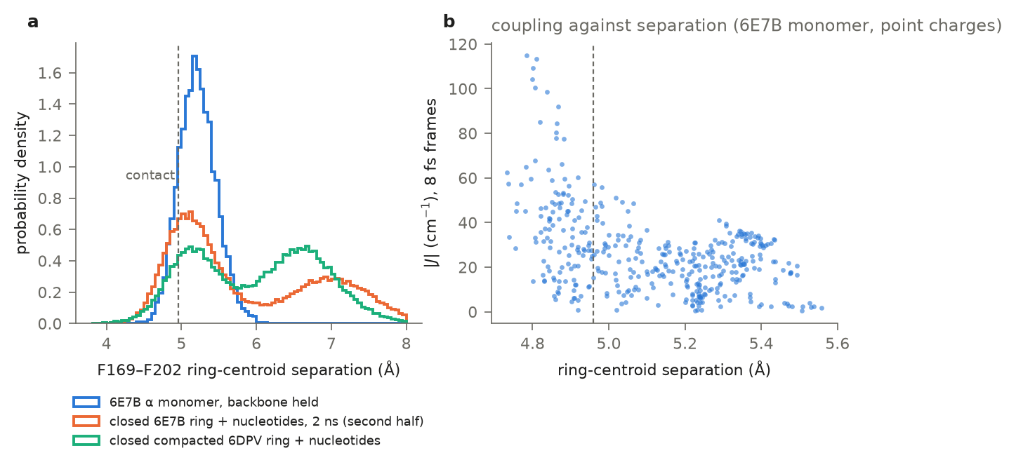
In the deposited models the ring centroids are 4.76 Å apart (6E7B) and 4.63–4.79 Å in all four α chains of the in situ axonal lattice [S1e]. In molecular dynamics of the α monomer the contact (< 4.96 Å) is one of two substates: 10–36% of the time across eight ff14SB trajectories and 34–35% with ff19SB/OPC and CHARMM36m [S1], in episodes of about one beat period (median 148–200 fs) [S1r]. Contact frames are strongly coupled (median 84 cm−1) and, with point-charge site energies, 76% of them strongly mixed ( > 0.5), against 19% of open frames [S1, S1ax]; with Cys200 in the quantum shell the native pair is detuned (Section 3.3). Coordinate uncertainty alone moves the deposited coupling about twofold either way [S1t], so the distributions, not single structures, carry the analysis. The maps themselves support the contact: the side chains are resolved (side-chain to backbone density 0.5–1.0), and in every contact entry the rings’ density centroids sit 0.05–0.27 Å further apart than the built contact, in both 6E7B half maps, while the compacted entry’s sit slightly closer; this is what a contact/open mixture averaged by the reconstruction would give [S1ah].
3.2 The lattice holds the contact, and compaction opens it
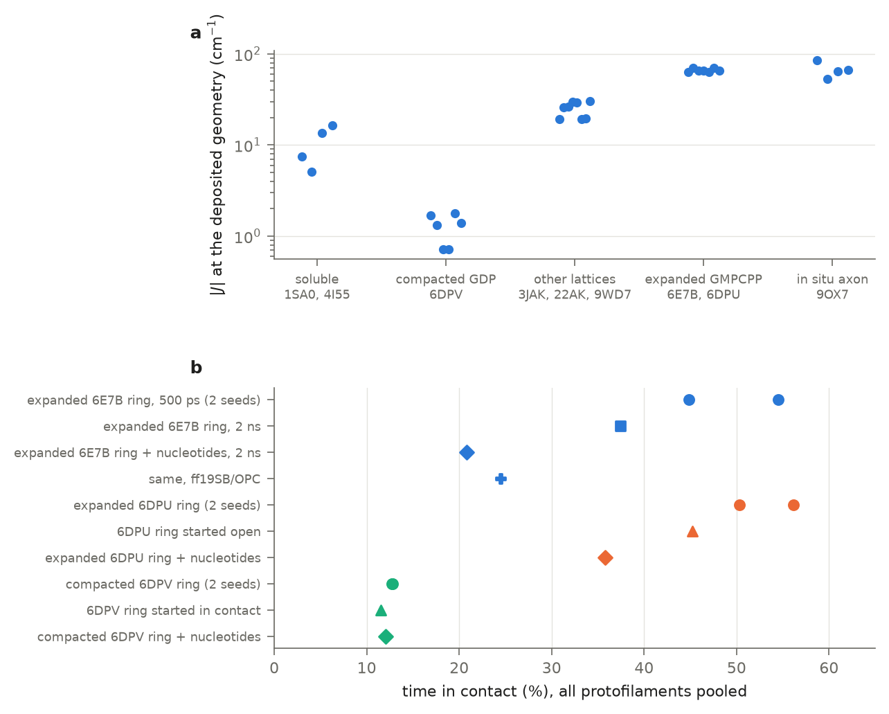
At the deposited geometries the coupling is 53–85 cm−1 in the in situ lattice, 63–70 in GMPCPP lattices, 19–30 in some lattice entries, 5–16 in soluble tubulin and 0.7–1.8 in the compacted GDP lattice 6DPV, where the pair is open (5.38 Å, 62°): about fifty-fold between the deposited expanded and compacted poses, following compaction rather than the nucleotide (9OX7 is GDP-bound but expanded) [S1m]. That contrast compares a contact pose with an open one; the coupling at a contact visited by a compacted lattice has not been computed. The structural switch matches the one reported for F202’s other neighbour, F255, which is parallel displaced in expanded lattices and T-shaped in compacted ones (Zehr et al., 2026); in the deposited models F255 sits 6.2 Å from F202 in both and couples to it at 0–11 cm−1 (to F169 at 0.5–2.5): a sixth or less of the pair’s own coupling where the pair is in contact, but more than it in the open compacted 6DPV (7.0 against 1.7 cm−1), where both are weak [S1an].
Dynamics give the same direction. Free isolated pieces lose the contact: the α monomer drifts from 37% to 9% contact, αβ dimers open it in three of three runs, and a four-chain patch holds it for about 300 ps before opening [S1, S1ad, S1ae]. Closed rings hold it. Over the first 500 ps, two 6E7B rings hold the contact 55% and 45% of the time and two expanded 6DPU rings 56% and 50%, against 12–13% in three compacted 6DPV rings, with or without nucleotides [S1af]. The expanded value is an early-time one: a 2 ns 6E7B ring with empty nucleotide pockets falls from 46% in its first fifth to 29% in its last; with GTP and Mg2+ bound in α and GMPCPP and Mg2+ in β it holds 21% overall, 29% in its first fifth and 13–21% across its second half; the same ring in ff19SB/OPC holds 24.5% (26% and 25% in its first and last fifths), although that force field doubles the monomer’s contact (15% to 34%) [S1af, S1]. A second expanded lattice, the nucleotide-bound 6DPU ring, holds 36% over 500 ps, falling from 42% in its first fifth to 21% in its last [S1af]. About a fifth to a quarter of the time is therefore the closed expanded lattice’s contact population in both force fields, against 12–13% for the compacted lattice, a contrast of about twofold (1.6–2.1) that holds for any contact threshold from 4.76 to 5.16 Å (1.6–2.3) [S1ao]; the expanded rings had not all stopped falling, and the compacted nucleotide-bound ring also fell (21% to 13% by fifths over 500 ps), so this is a finite-trajectory estimate whose direction under longer sampling is not known, not a converged value. Swapping the starting pose does not change the contact: a compacted ring started with the expanded lattice’s contact reopens to 12%, and an expanded ring started with the compacted lattice’s open pose returns to 45% [S1af]. Within one ring the protofilaments range from 0 to 90% contact, and they share one lattice, so each ring is one sample.
3.3 Cys200 sets the detuning
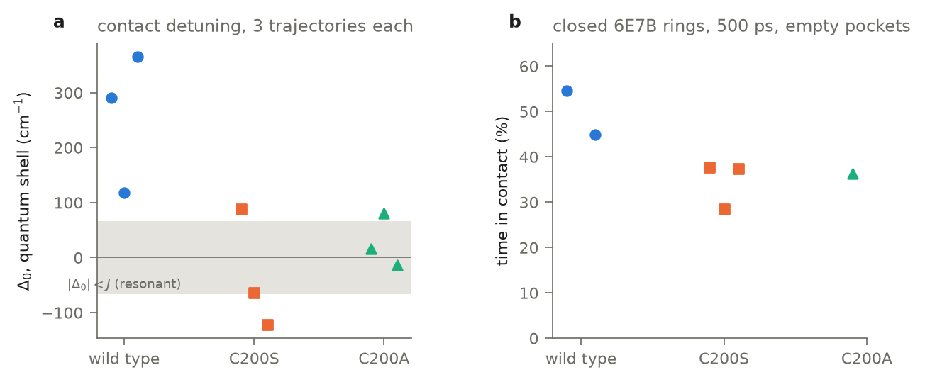
With the nearest residues treated quantum mechanically, the wild-type contact is detuned by +257 ± 73 cm−1 (mean and standard error over three trajectories of six contact frames each; +117 to +365 per trajectory) [S1au, S1ax], F202 pushed down on the side the Cys200 thiol touches [S1]. Replacing the cysteine removes most of that detuning, by either substitution: C200S gives −32 ± 63 cm−1 (+89, −63, −122) and C200A +28 ± 27 (+17, +80, −13), close to removing Cys200 from the quantum shell computationally. The two mutants are not distinguishable (0.9 standard errors) and each lies about three standard errors from the wild type; with six frames per trajectory the individual trajectories scatter by about ±100 cm−1 around the coupling scale [S1g, S1au, S1ax]. Neither mutation disturbs the pair geometry [S1g]. The thiol itself flickers rather than gates: its S-H points at F202 95% of the time with excursions of tens of femtoseconds [S1s].
3.4 What the coupling noise is made of
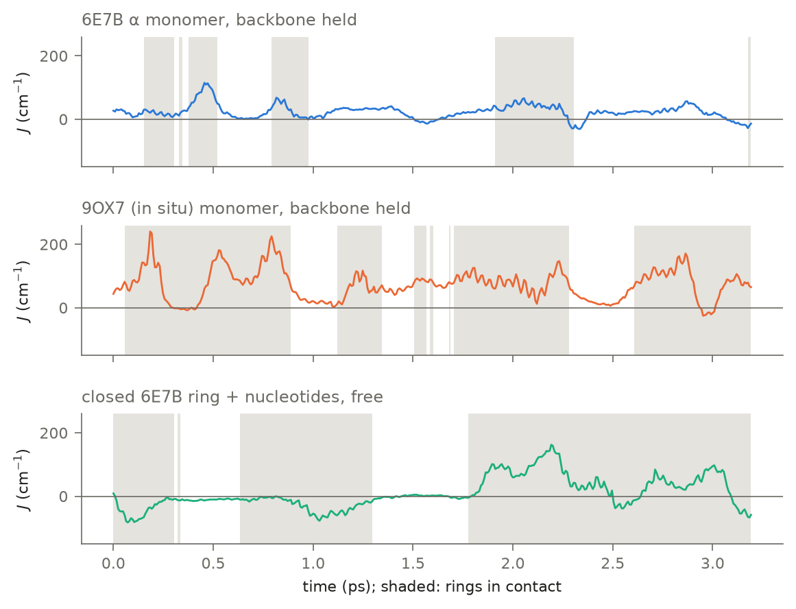
On consecutive 8 fs frames the coupling fluctuates by 65% of its magnitude at the contact, 74–78% with the sign kept, with a memory of 72–80 fs, and changes sign 13 times in 3.2 ps [S1l, S1aa]. Started from the in situ pose (9OX7) the pair is 20–30% quieter (0.55 and 0.57, four to six sign changes) [S1ag]. In the closed lattice ring it is larger: the contact coupling is weaker (28–39 cm−1), its signed fluctuation 1.28–1.32 of its magnitude, the minority sign held 30–38% of the time and the memory 112–160 fs; with nucleotides bound it is 1.06–1.21 at 37–40 cm−1 (minority sign 36–44%, memory 176–272 fs) [S1aa, S1aj]. The ring separation explains only 13% of it; an in-plane map of J reproduces the directly computed noise from the MD’s slip and rotation, which places the deposited pose on a steep slope beside a line where Coulomb and exchange cancel [S1l, S1ac]. About one angstrom of slip away the coupling has a maximum, but the rings do not become quieter there: computed directly, the coupling roughly doubles while its relative noise falls by only about a fifth and its sign still changes, and reaching it costs 2.8 ± 0.4 [S1ac]. The gap noise has a 56 fs memory, a fast quarter above 400 cm−1, and nearly independent sites [S1j]; the first shell dominates the wild type’s [S1]. Charge transfer stays a minor part of the lowest pair (1–6%), and the extra gap noise from its difference dipole is small (about 8–10 cm−1) [S1i]. Diabatizing the two lowest states of the full two-ring calculation separates what the fragment picture misses: a coupling 1.3–1.5 times larger, by the same factor in open and contact geometries (so not a charge-transfer effect), an extra detuning of 30–50 cm−1, and at the contact a geometry-driven detuning fluctuation of about 35 cm−1 [S1i]. These move the coherence numbers by less than their spread (Section 3.5). A larger basis with diffuse functions (def2-SVPD) scales the fragment coupling by 2.2 (frame-to-frame correlation 0.994) and leaves its relative noise where it was (0.75 against 0.77); diabatized in that basis the supermolecular coupling is 0.72–0.88 of the fragment value [S1ai]. Computed directly on frames of the nucleotide-bound lattice, the factor is smaller there: 1.97 for the mean signed contact coupling and 1.87 for its standard deviation (frame correlation 0.98–0.999, the sign agreeing on every frame) [S1ap].
3.5 Coherence compared with FMO
Each environment is described by its own 8 fs record: the static coupling is the mean signed coupling over the record’s contact frames, the coupling noise is its standard deviation over the same frames, with the record’s own memory, and the site energies are the contact-frame values of Section 3.3, treated five ways [S1ak] (Table S3). Lifetimes are given in femtoseconds and against the FMO BChl 1–2 pair computed with the same code and detector, whose eigenstate coherence lasts 80 fs at 300 K (0.51 of its 157 fs beat) and whose visible population beats last 377 fs [S1u, S1ak].
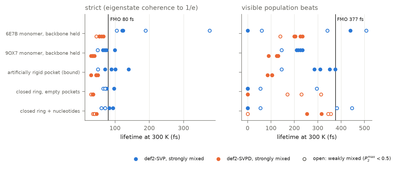
In the closed, nucleotide-bound lattice the mean signed coupling is 22 cm−1 (43 cm−1 in def2-SVPD, with the basis factor measured on lattice frames), against a standard deviation of 52 (98): the coupling’s noise exceeds its mean. Whether the pair mixes then depends on Cys200. With the cysteine in the quantum shell the native pair stays localized ( 0.02–0.10); with Cys200 left out of the quantum shell, or replaced by alanine, it mixes almost fully (0.87–0.99); with serine, or with point-charge site energies, it is weakly mixed in def2-SVP (0.19) and about half mixed in def2-SVPD (0.48–0.53). Which corner is strongly mixed moves with the ring-geometry treatment; that one is (Section 3.3) [S1ak, S1ap, S1ax]. Where it is strongly mixed, the pair keeps eigenstate coherence for 39–94 fs at 300 K, the same order as FMO’s 80 fs, under every ring-geometry treatment; because its beat is slower (period 260–760 fs), that is about a tenth of an oscillation against FMO’s half [S1ak, S1ap, S1ax]. With the monomer’s basis factor carried to the lattice instead, the def2-SVPD rows give 35–36 fs [S1ak]. Visible population beats are not robust here. At the strongly mixed corners they range from none to about 0.4 ps, and the same corner moves across that range with the hierarchy depth (Cys200 left out, def2-SVPD: 437, 391, 316, 199, 0 and 0 fs at depth 4 to 9), with the detector’s threshold (from 0.1% to 2% of the population, 3.4 periods to none) and with the basis; FMO’s visible beats stay at 370–377 fs at every depth, and the eigenstate lifetime of both systems does not move [S1al, S1aq]. The lattice pair’s population oscillation sits at the detector’s limit, so we read no lifetime from it. With empty nucleotide pockets the lattice is noisier, the strongly mixed corners keep 35–98 fs and no beat is visible [S1ak].
What sets the eigenstate lifetime is the coupling noise. Switching the gap bath off leaves it unchanged with Cys200 left out and shortens it by about a fifth for C200A, while switching the coupling bath off lengthens it 2.6- to 3.9-fold for both [S1ar]. It is therefore a dephasing time, nearly the same for a pair that barely mixes (60 fs at 0.02, def2-SVP) as for one that mixes fully (84 fs at 0.97), when both baths carry the coupling record’s memory, as in every row of Table S3. The mixing shows instead in how much population crosses to the other ring before the coherence is lost: 0.30–0.36 for the strongly mixed corners against 0.13–0.21 for the native full shell [S1ar]. Both statements rest on that shared memory for the native pair in def2-SVP: given its own measured memory (56 fs [S1j], against the coupling’s 224 fs), the gap noise leaves every strongly mixed lifetime and crossed population unchanged, but the barely mixed native shell then keeps its eigenstate coherence for 281 fs and passes 0.42 of the population by then, driven by the gap noise rather than by mixing; in def2-SVPD it is unchanged (35 fs, 0.21) [S1ba]. The FMO reference carries no coupling bath; given one of the Phe lattice’s relative size (standard deviation 1.0 and 2.4 times the coupling), its eigenstate lifetime falls from 80 to 41 and 20 fs [S1ar], so the comparison with FMO is a comparison of dephasing times under different baths.
The other environments show what moves it; only the coupling bath changes between them, the gap bath being the monomer’s contact-frame bath throughout. In the free monomer pocket, where the coupling is larger than its noise, strongly mixed pairs keep 54–124 fs (visible beats to 440 fs); started from the in situ structure, 30–100 fs; in a pocket held rigid at the cryo-EM pose (every heavy atom within 8 Å held), 30–140 fs [S1ak]. In absolute time the lattice therefore shortens the coherence by about 1.3–1.7 times against the free monomer, corner for corner, and the rigid pocket does not lengthen it, because its larger coupling shortens the beat as much as it narrows the noise. The coupling memory (176 or 272 fs, the two protofilaments) leaves the eigenstate lifetime unchanged and moves the visible beats; cooling with the 300 K noise amplitudes kept leaves it unchanged, and scaling both noise amplitudes by the square root of the temperature lengthens it by 14–38% at 150 K for the strongly mixed corners [S1al]. Contact-open switching, the ring-breathing mode and the gap-coupling correlation do not change the picture [S1s, S1h, S1w, S1aa]. A vibration at the exciton splitting would lengthen visible beats by 40–50% without lengthening the electronic coherence, the same ambiguity that marked the FMO debate [S1z].
3.6 Experimental tests, and what would refute each
Each prediction is stated with the result that would support it, the result that would refute it, and the design that makes the phenylalanine signal visible at all. That last part is the practical crux: Phe absorbs far more weakly than Trp and sits under the Tyr and Trp bands (Mihalyi, 1968; Pace et al., 1995), so every test below is a difference between variants (F169L, F202L, C200S, C200A, wild type) or between lattice states measured on the same sample, never an absolute spectrum. These are in vitro tests on purified microtubules; the in situ structure (9OX7) is what justifies starting from expanded lattices [S1e].
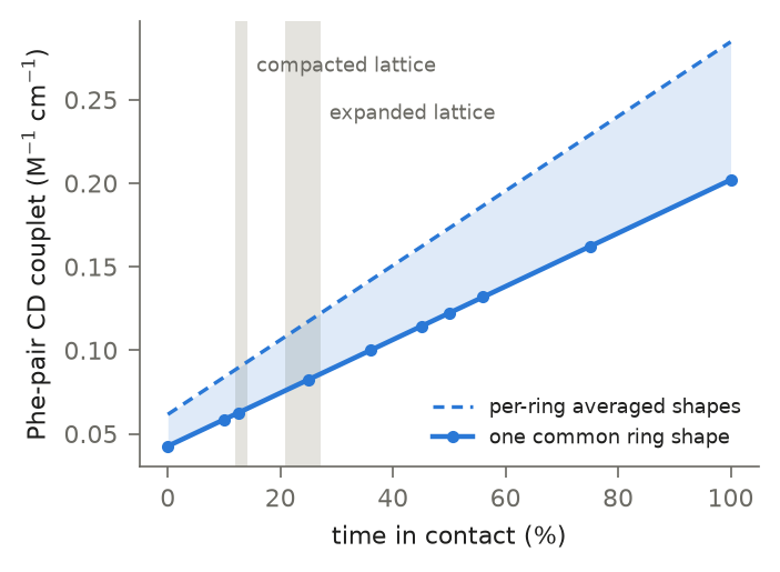
(1) The lattice switch. Prediction: expanded lattices (GMPCPP, taxol-stabilized) hold the contact about a fifth to a quarter of the time and, at the deposited geometries, couple the pair at 53–85 cm−1; compacted GDP lattices hold it about an eighth of the time and couple it at ~1 cm−1 in the deposited open pose; the coupling at a contact visited by a compacted lattice was not computed [S1m, S1af]. Near-UV CD, computed by the exciton matrix method (Bayley et al., 1969), reads the contact and the coupling [S1k], and the pair’s couplet rises with the contact fraction (0.04 to 0.28 M−1 cm−1 across the contact range; the amplitude depends on the ring geometry at the milliångström level through the direction of the weak Phe transition dipole, so it is given as a range over two ring-geometry treatments) [S1, S1ay]. Supports: a Phe-pair couplet (wild type minus F169L or F202L) larger in expanded than in compacted lattices by about the predicted amount (0.076–0.122 against 0.062–0.093 M−1 cm−1, higher in every pairing of simulation seeds and ring-geometry treatments, by 16–39%; not an on/off switch). Refutes: no change in the difference couplet between lattice states, or a change that does not track compaction (for example GDP-bound but expanded lattices behaving like compacted ones). Design: the same tubulin polymerized with GMPCPP and with GDP (with and without taxol, with end-binding proteins that favour compaction, and decorated with kinesin in its strong-binding state, reported to expand GDP lattices; Peet et al., 2018), each as wild type and F169L; the Phe-pair signal is the wild-type minus F169L difference. Each mutant needs matched polymer mass, protofilament number and lattice spacing, and a structural or spectroscopic check that it keeps the pocket and the compaction response. A lattice-expanding treatment should raise the signal and a compacting one lower it. Read at the closed rings’ own contact fractions (2 ns, two seeds each: compacted 12.0–14.2%, expanded 20.8–27.2%), the couplet is 0.062–0.093 M−1 cm−1 for the compacted lattice and 0.076–0.122 for the expanded one, a difference of 0.011–0.034 M−1 cm−1 from the contact population alone [S1am, S1ay]; a measurement must resolve a mutant-difference couplet to that precision, on polymerized microtubules, with light scattering and the Tyr/Trp background removed by the difference.
(2) The Cys200 gate. Prediction: replacing Cys200, by serine or by alanine, removes most of the native detuning ( within about ±120 cm−1 in every mutant trajectory, against +117 to +365 in the wild type) [S1g, S1au, S1ax]; in 500 ps rings with empty nucleotide pockets the closed lattice holds the C200S contact about a third less than the wild type (28–38% over three rings, against 45–55%) [S1af]. CD is not the test of this: it reads the contact and the coupling rather than the mixing, and its amplitude depends on the ring geometry [S1k, S1ay]. Supports: the F169 and F202 site energies, read from single-ring difference absorption (F169L and F202L, each in wild-type and C200S backgrounds) or from low-temperature 0–0 bands, converge in C200S and C200A and stay apart in the wild type. Refutes: mutant site energies as far apart as the wild type’s. Design: four single mutants and two double mutants in two backgrounds; difference spectra at low temperature, where the 0–0 bands sharpen.
(3) Coherence. Prediction: in contact in an expanded lattice, a strongly mixed pair (with the native detuning removed: Cys200 replaced, or left out of the quantum shell) keeps eigenstate coherence for 39–94 fs at 300 K (the strongly mixed nucleotide-bound lattice rows of Table S3), the same order as FMO’s 80 fs with the same machinery and detector and about a tenth of its 260–760 fs beat; whether a visible population beat survives is beyond what the model resolves, and the native pair, detuned by Cys200, barely mixes [S1ak]. A vibration near the exciton splitting would lengthen visible beats by 40–50% without lengthening the electronic coherence [S1z]. Supports: a cross peak oscillating at the exciton splitting in the Phe-pair difference signal, damping within about one period, stronger in C200S than in the wild type, present in expanded lattices and absent in compacted ones and in F169L. Refutes (that the beat is electronic): an oscillation whose frequency shifts with ring deuteration or does not change with lattice state. Refutes (the model’s pessimism): beats in the difference signal lasting several periods at physiological temperature. Design: two-dimensional electronic spectroscopy (or pump-probe) near 260 nm on polymerized microtubules, wild type minus F169L, expanded vs compacted lattice, with a deuterated-ring or temperature series to separate vibronic from electronic beats. This is the hardest measurement here and should follow tests (1) and (2).
4. Discussion
A state the microtubule enters
The coupled pair is not a fixed property of tubulin. In closed lattices the contact is present about a fifth to a quarter of the time when the lattice is expanded and about an eighth of the time when it is compacted, and the in situ axonal lattice, GDP-bound but expanded, holds it [S1e, S1m, S1af]. The same compaction that opens this contact flips F202’s other neighbour, F255, from parallel-displaced to T-shaped, in the relay that carries the effect of GTP hydrolysis from the longitudinal to the lateral interface (Zehr et al., 2026). The exciton is therefore a state the lattice enters or leaves, set by its spacing rather than by the nucleotide alone, and it sits on a structural pathway the lattice already uses.
In a cell that spacing is not uniform. The GTP cap at growing ends is thought to be expanded and the GDP lattice behind it compacted (Alushin et al., 2014; Zhang et al., 2015); kinesin in its strong-binding state has been reported to expand GDP lattices locally (Peet et al., 2018; Shima et al., 2018); microtubule-associated proteins, end-binding proteins and stabilizing drugs such as taxol favour one state or the other (Alushin et al., 2014; Siahaan et al., 2022; Zhang et al., 2015). Stable axonal microtubules appear expanded in situ (Zehr et al., 2026). If these hold, the pair is coupled near growing tips, along stretches under motor traffic and in stabilized axonal microtubules, and largely off along compacted dynamic shafts. The question the warm-wet argument asks then becomes conditional and answerable: in which microtubules, and when, is the pair coupled? We have simulated the two endpoints, expanded and compacted, not a motor- or MAP-bound lattice or the cap-to-shaft transition; those are the direct next tests.
How long the coherence lasts, and what limits it
The warm-wet argument expects electronic coherence in a protein at physiological temperature to be gone faster than anything that matters. In this pair, where it is strongly mixed, the eigenstate coherence lasts 39–94 fs in the closed, nucleotide-bound lattice and up to 124 fs in a free monomer pocket, against 80 fs for FMO’s BChl 1–2 pair computed with the same machinery and detector [S1ak, S1ap]. That is the timescale of the system usually cited as coherent at room temperature, not a vanishing one, although FMO’s would be shorter still under coupling noise of the Phe lattice’s relative size [S1ar]. In both it is a dephasing time; what distinguishes the coupled pair from a detuned one is not how long the coherence lasts but how much population crosses before it is lost [S1ar], with the caveat that a faster gap bath lets the detuned native pair exchange population through the noise alone [S1ba]. It is also a fraction of one oscillation of this slow pair, and a visible population beat in the lattice cannot be resolved robustly: it comes and goes with the basis set, the detection threshold and the hierarchy depth [S1ak, S1al, S1aq]. Two conditions gate it. The lattice must hold the contact, which happens about a fifth to a quarter of the time in the expanded lattice [S1af]. And the pair must be in resonance: with Cys200 treated quantum mechanically the native pair is detuned and barely mixes, so the strongly mixed exciton belongs to the pair with Cys200 removed, by either substitution, or to the native pair only under simpler descriptions of its environment [S1g, S1ak, S1ax].
What limits it is computed, not assumed. The coupling noise sets the ceiling: the rings slide across a steep coupling slope beside a line where Coulomb and exchange cancel, so in the lattice the coupling fluctuates by more than its mean and changes sign [S1l, S1ac, S1aj, S1ak]. Each condition moves the lifetime by a computed amount: the closed lattice shortens it about 1.3–1.7 times against the free monomer; a pocket held rigid does not lengthen it in absolute time; nucleotides quiet the lattice noise by 10–25%; cooling lengthens it by 14–38% at 150 K only if the noise falls with temperature; the coupling memory, two-state switching, the ring-breathing mode, charge transfer and the gap-coupling correlation do not move it [S1ak, S1al, S1n, S1s, S1h, S1i, S1w]. Read together, these are a map of what it would take: an aromatic contact whose surroundings hold the rings against slip and whose sites are in resonance should keep its coherence longer. Such contacts may exist in more rigid environments than this pocket, in tightly packed sheets, ligand pockets or engineered variants, and a census of aromatic contacts ranked by the rigidity of their environment and the steepness of their coupling slope would show where this pair sits and where stronger cases should be looked for.
Does it matter?
Whether coherence of this length matters is a separate question from whether it occurs, with two parts.
Here. We do not show a function, and the treatment cannot. What it shows is what the state could do. In a toy model with the contact held, infinite-temperature dephasing and an artificial trap, the noise that cuts the beat makes excitation transfer between the rings nearly as fast as any dephasing allows: within 0–7% of the optimum, about six times faster than coherent transfer in the detuned wild type [S1x], the environment-assisted transport regime described for light-harvesting networks (Plenio & Huelga, 2008; Rebentrost et al., 2009). That transfer takes about 2 ps, much longer than a contact episode (about 150 fs), so in the lattice it would be interrupted many times; the rate holds only while the contact does. If phenylalanine excitation is handed on to nearby tyrosine and tryptophan, as is common in proteins (Macmillan et al., 2008; Searcy et al., 1989), this pair is better suited to moving energy than to holding a coherent wave. The coupled state is also regulated: the lattice switches it with compaction, and in a cell compaction varies with growth, motor traffic and stabilization (above). A structure that turns an excitonic state on and off through processes it already undergoes has the kind of property a functional mechanism would need; having the property is not evidence that one exists. The excitation source is a real constraint: phenylalanine absorbs near 257 nm, which does not reach neurons as light, and the internal source proposed for cells, excited states made by oxidative chemistry, emits at 350 nm and longer (Pospíšil et al., 2014), below the energy needed to excite phenylalanine by light or by direct transfer. How the pair would be excited in a cell is an open question this work does not answer.
A possible non-optical route. That constraint applies to the singlet studied here. Phenylalanine’s lowest triplet lies lower: its relaxed (0–0) energy, computed directly at four levels of theory, is 3.48–3.52 eV, and the same method places benzene’s triplet within 0.07 eV of its measured 3.66 eV [S1av]. That is at the top of the energy range of the triplet carbonyls oxidative chemistry produces (Pospíšil et al., 2014), so transfer from the most energetic of them would be roughly thermoneutral, and uphill from most. Triplet transfer is an exchange process and needs the kind of van der Waals contact this pair is built on; aromatic triplet interactions in proteins likewise depend on a stacked contact, which a folded structure usually prevents (Gonnelli & Strambini, 2005) and which F169/F202 has. With the Cys200 quantum shell, the contact shares a triplet on 10 of 18 contact frames, at least as often as it shares the singlet computed the same way (8 of 18) [S1av]. Two conditions decide whether the route operates. First, the rings are buried: no carbonyl-sized molecule reaches F169, F202 or their phenylalanine neighbours F138 and F255 in four deposited structures [S1aw], so a donor would have to form in contact, for example by oxidation of a contacting residue, or reach the cluster during a transient opening; the same burial shields a triplet from oxygen. Second, the step is at best thermoneutral. We record this as a hypothesis the present results make testable, not as a result.
FMO as a scale
The comparison with FMO is a scale, not a claim of equivalence. Long-lived FMO beats were first read as electronic coherence (Engel et al., 2007), and later work assigns much of that beating to vibrational and vibronic motion (Duan et al., 2017). The FMO pair computed here therefore places the phenylalanine pair against the system that defined the question, with the same code and detector [S1u, S1ak]; our detector is stricter than the published visibility criterion, so the comparison is internal. It does not say that either system uses coherence. The same vibronic ambiguity applies here: a vibration at the pair’s splitting would lengthen its visible beats without lengthening its electronic coherence [S1z], so a measured beat would need an isotope or temperature test of its frequency before it is read as electronic.
Limits of the treatment
The couplings and site energies are fragment TDDFT at wB97X/def2-SVP, with rigid rings (no internal vibrations) and point-charge embedding without polarization. Both rings carry one averaged ring geometry, each residue keeping its own side-chain anchor: averaging each ring’s own MD geometry, as version 1.0 did, injected a trajectory-dependent detuning of tens to ~180 cm−1, and the detunings, mixing and CD of this version are recomputed without it [S1ax]. The CD amplitude remains uncertain by about a factor of two, because the direction of the weak Phe transition dipole follows the ring geometry at the milliångström level [S1ay]. A third phenylalanine, F138 (5.4 Å from F169), couples to F169 in the singlet at −1.8 ± 25 cm−1 and lies about 450 cm−1 below it, so it never mixes coherently into the pair ( at most 0.08), and the two-site treatment stands [S1az]. The supermolecular diabatization corrects the coupling and adds a detuning on a subset of frames [S1aa]. The larger def2-SVPD basis scales the coupling by 2.2 in the monomer (100 frames) and by 1.97, its noise by 1.87, in the nucleotide-bound lattice, measured on the first 56 frames of each of two protofilaments; the other environments use the monomer factor [S1ai, S1ap]. The baths are classical and Gaussian. Driving the pair with the recorded coupling series itself instead gives the same eigenstate lifetime within a factor of 0.92–1.24 over windows at least 80% in contact, and a classical Gaussian drive reproduces the HEOM within 0.90–1.05 at the same bath memories [S1as]; the supermolecular (diabatized) coupling in place of the fragment one keeps the strongly mixed lattice pairs at 48–67 fs [S1at]. The static coupling and its noise are the mean and standard deviation of the signed coupling over contact frames (17–27 and 48–55 cm−1 for contact thresholds of 4.76–5.16 Å [S1ao]), with a memory taken over the whole record (224 fs; 248 fs within contact episodes, which leaves the eigenstate lifetime unchanged [S1aq]), and the gap bath uses the monomer’s contact-frame site energies in every environment [S1ak]; switching it off changes the strongly mixed lifetimes by at most a fifth and lengthens only the barely mixed full shell’s [S1ar]; it carries the coupling record’s memory, not its own measured 56 fs, which leaves the strongly mixed rows unchanged but not the native shell in def2-SVP [S1ba]; and the lattice’s own site energies were not computed. The visible-beat measure is not robust in the lattice (threshold, basis and hierarchy depth) and carries no conclusion; the eigenstate lifetime is converged in hierarchy depth and Matsubara terms [S1al, S1aq]. The model is a two-site pair. F202’s second neighbour F255 couples to it at 0–11 cm−1 at the deposited geometries and to F169 at 0.5–2.5 [S1an]. A coupling of 10 cm−1 exchanges energy in about 0.8 ps, ten to twenty times longer than the pair’s 39–94 fs coherence, so it leaves the coherence numbers unchanged; on the picosecond scale of transfer it is a competing channel, and it is left out of the transfer model too. Its coupling along the dynamics and its site energy were not computed. The tyrosine and tryptophan acceptors that would receive transferred energy are not in the model either. The dynamics run 500 ps to 2 ns, in ff14SB and, for the nucleotide-bound ring, ff19SB/OPC; in the monomer ff14SB opens this pair more than ff19SB/OPC or CHARMM36m do, but in the closed ring the two agree (21% and 24.5%) [S1, S1af]. The closed rings carry nucleotides but no C-terminal tails, MAPs, taxol or axial curvature. The lattice noise comes from two protofilaments of each of two rings, 3.2 ps each; the two nucleotide-bound windows were in contact 76% and 47% of the time, well above the ring’s 21% over 2 ns, and whether less contact-rich protofilaments carry different coupling statistics is not known [S1aa, S1aj]. Every quantity is computed; the experiments in Section 3.6 are what would measure them.
Data and code availability
The code, results and structures behind every number are deposited as a separate Zenodo software record, version 1.1.0 (https://doi.org/10.5281/zenodo.23990898; archive SHA-256 5bd18eae0a6ac7841d146400de6139fbb4f831d1fb1d2322e23f136f890b19c3; all versions: https://doi.org/10.5281/zenodo.23858681). It holds every producer cited through the bracketed keys (Table S0) with the local modules they import, the result files they wrote, the ring and pair trajectories the figures read, the nucleotide parameters, the deposited structures used, the figure script and the topology and operator tests. Its SHA-256 manifest is checked, and the figures are redrawn, from a fresh extraction before release. The 8 fs coordinate dumps (about 7 MB each) are not included; each is regenerated by its producer with the recorded seed. Deposited structures are from the wwPDB (6E7B, 6DPU, 6DPV, 9OX7, 22AK, 3JAK, 9WD7, 1SA0, 4I55) and maps from EMDB (EMD-8997 with its half maps, EMD-7973, EMD-7974, EMD-70956).
This is version 1.1 of the paper (version 1.0: https://doi.org/10.5281/zenodo.23858971). It corrects the ring geometry behind every detuning, mixing and circular-dichroism number, gives the circular-dichroism readout as a range, and adds the third phenylalanine, the triplet and a check of the gap bath’s memory; Supplementary Section S12 lists the changes with the version 1.0 values, and the default mode of the archived producers still reproduces version 1.0.
Conflict of interest
The author is the founder and developer of Synthesis Lab, a commercial research-writing platform, and has a financial interest in it. Synthesis Lab was used in preparing this manuscript (see the Generative-AI disclosure); it is not a subject of the study, and every reported calculation is produced by the archived code, which runs without it.
Generative-AI disclosure
Generative-AI assistance was used in preparing this manuscript, its supplement and the code archive between 21 October and 1 November 2026 (the code from 21 October, the manuscript from 25 October). Anthropic Claude Opus 5.5, accessed through the Claude Code command-line interface, was used for code authorship and code review (the producers, the system builders and the test suite); launching, monitoring and analysing the calculations on the author’s own machines; the figure scripts; checking references against PubMed and Crossref; and drafting the manuscript and supplement. These model identities and dates are taken from the co-authorship trailers in the project’s version history; the code archive lists every commit behind its files with its date and trailer (AI_HISTORY.tsv). The oldest archived scripts date from August 2026; their commits name Claude Opus 5, accessed the same way, or no assistant. Synthesis Lab, a manuscript platform the author develops, was used to attach and format the references (citeSources) and to obtain simulated peer reviews of the first draft on 31 October 2026 and follow-up reviews of the revision on 1 November 2026: OpenAI GPT-5.6 Sol and Anthropic Claude Opus 5, both accessed through the OpenRouter API, each with a citation audit by Anthropic Claude Sonnet 5.5; their concerns, and the calculations they asked for, shaped this version.
No AI system is an author, and no text, number or figure entered the manuscript without author review. All calculations, sources, citations and conclusions were checked and approved by the author, who takes full responsibility for the content.
References
Adolphs, J., & Renger, T. (2006). How Proteins Trigger Excitation Energy Transfer in the FMO Complex of Green Sulfur Bacteria. Biophysical Journal, 91(8), 2778–2797. https://doi.org/10.1529/biophysj.105.079483
Alushin, G. M., Lander, G. C., Kellogg, E. H., Zhang, R., Baker, D., & Nogales, E. (2014). High-Resolution Microtubule Structures Reveal the Structural Transitions in αβ-Tubulin upon GTP Hydrolysis. Cell, 157(5), 1117–1129. https://doi.org/10.1016/j.cell.2014.03.053
Babcock, N. S., Montes-Cabrera, G., Oberhofer, K. E., Chergui, M., Celardo, G. L., & Kurian, P. (2024). Ultraviolet superradiance from mega-networks of tryptophan in biological architectures. The Journal of Physical Chemistry B, 128(17), 4035–4046. https://doi.org/10.1021/acs.jpcb.3c07936
Bayley, P. M., Nielsen, E. B., & Schellman, J. A. (1969). Rotatory properties of molecules containing two peptide groups: theory. The Journal of Physical Chemistry, 73(1), 228–243. https://doi.org/10.1021/j100721a038
Chai, J.-D., & Head-Gordon, M. (2008). Systematic optimization of long-range corrected hybrid density functionals. The Journal of Chemical Physics, 128(8), Article 084106. https://doi.org/10.1063/1.2834918
Duan, H.-G., Prokhorenko, V. I., Cogdell, R. J., Ashraf, K., Stevens, A. L., Thorwart, M., & Miller, R. J. D. (2017). Nature does not rely on long-lived electronic quantum coherence for photosynthetic energy transfer. Proceedings of the National Academy of Sciences, 114(32), 8493–8498. https://doi.org/10.1073/pnas.1702261114
Eastman, P., Galvelis, R., Peláez, R. P., Abreu, C. R. A., Farr, S. E., Gallicchio, E., Gorenko, A., Henry, M. M., Hu, F., Huang, J., Krämer, A., Michel, J., Mitchell, J. A., Pande, V. S., Rodrigues, J. P., Rodriguez-Guerra, J., Simmonett, A. C., Singh, S., Swails, J., … Markland, T. E. (2024). OpenMM 8: Molecular Dynamics Simulation with Machine Learning Potentials. The Journal of Physical Chemistry B, 128(1), 109–116. https://doi.org/10.1021/acs.jpcb.3c06662
Eastman, P., Swails, J., Chodera, J. D., McGibbon, R. T., Zhao, Y., Beauchamp, K. A., Wang, L.-P., Simmonett, A. C., Harrigan, M. P., Stern, C. D., Wiewiora, R. P., Brooks, B. R., & Pande, V. S. (2017). OpenMM 7: Rapid development of high performance algorithms for molecular dynamics. PLOS Computational Biology, 13(7), Article e1005659. https://doi.org/10.1371/journal.pcbi.1005659
Engel, G. S., Calhoun, T. R., Read, E. L., Ahn, T.-K., Mančal, T., Cheng, Y.-C., Blankenship, R. E., & Fleming, G. R. (2007). Evidence for wavelike energy transfer through quantum coherence in photosynthetic systems. Nature, 446(7137), 782–786. https://doi.org/10.1038/nature05678
Gonnelli, M., & Strambini, G. B. (2005). Intramolecular quenching of tryptophan phosphorescence in short peptides and proteins. Photochemistry and Photobiology, 81(3), 614–622. https://doi.org/10.1562/2004-11-09-RA-367
Hameroff, S., & Penrose, R. (2014). Consciousness in the universe: A review of the ‘Orch OR’ theory. Physics of Life Reviews, 11(1), 39–78. https://doi.org/10.1016/j.plrev.2013.08.002
Huang, J., Rauscher, S., Nawrocki, G., Ran, T., Feig, M., de Groot, B. L., Grubmüller, H., & MacKerell, A. D. (2017). CHARMM36m: an improved force field for folded and intrinsically disordered proteins. Nature Methods, 14(1), 71–73. https://doi.org/10.1038/nmeth.4067
Ishizaki, A., & Fleming, G. R. (2009). Theoretical examination of quantum coherence in a photosynthetic system at physiological temperature. Proceedings of the National Academy of Sciences, 106(41), 17255–17260. https://doi.org/10.1073/pnas.0908989106
Izadi, S., Anandakrishnan, R., & Onufriev, A. V. (2014). Building Water Models: A Different Approach. The Journal of Physical Chemistry Letters, 5(21), 3863–3871. https://doi.org/10.1021/jz501780a
Jakalian, A., Jack, D. B., & Bayly, C. I. (2002). Fast, efficient generation of high‐quality atomic charges. AM1‐BCC model: II. Parameterization and validation. Journal of Computational Chemistry, 23(16), 1623–1641. https://doi.org/10.1002/jcc.10128
Johansson, J., Nation, P., & Nori, F. (2013). QuTiP 2: A Python framework for the dynamics of open quantum systems. Computer Physics Communications, 184(4), 1234–1240. https://doi.org/10.1016/j.cpc.2012.11.019
Jorgensen, W. L., Chandrasekhar, J., Madura, J. D., Impey, R. W., & Klein, M. L. (1983). Comparison of simple potential functions for simulating liquid water. The Journal of Chemical Physics, 79(2), 926–935. https://doi.org/10.1063/1.445869
Kalra, A. P., Benny, A., Travis, S. M., Zizzi, E. A., Morales-Sanchez, A., Oblinsky, D. G., Craddock, T. J. A., Hameroff, S. R., MacIver, M. B., Tuszyński, J. A., Petry, S., Penrose, R., & Scholes, G. D. (2023). Electronic Energy Migration in Microtubules. ACS Central Science, 9(3), 352–361. https://doi.org/10.1021/acscentsci.2c01114
Lambert, N., Raheja, T., Cross, S., Menczel, P., Ahmed, S., Pitchford, A., Burgarth, D., & Nori, F. (2023). QuTiP-BoFiN: A bosonic and fermionic numerical hierarchical-equations-of-motion library with applications in light-harvesting, quantum control, and single-molecule electronics. Physical Review Research, 5(1), Article 013181. https://doi.org/10.1103/physrevresearch.5.013181
Li, R., Sun, Q., Zhang, X., & Chan, G. K.-L. (2025). Introducing GPU Acceleration into the Python-Based Simulations of Chemistry Framework. The Journal of Physical Chemistry A, 129(5), 1459–1468. https://doi.org/10.1021/acs.jpca.4c05876
Macmillan, A. M., McGuinness, C. D., Sagoo, K., McLoskey, D., Pickup, J. C., & Birch, D. J. S. (2008). Effect of pH on Aqueous Phenylalanine Studied Using a 265‐nm Pulsed Light‐emitting Diode. Annals of the New York Academy of Sciences, 1130(1), 300–304. https://doi.org/10.1196/annals.1430.056
Maier, J. A., Martinez, C., Kasavajhala, K., Wickstrom, L., Hauser, K. E., & Simmerling, C. (2015). ff14SB: Improving the Accuracy of Protein Side Chain and Backbone Parameters from ff99SB. Journal of Chemical Theory and Computation, 11(8), 3696–3713. https://doi.org/10.1021/acs.jctc.5b00255
McKemmish, L. K., Reimers, J. R., McKenzie, R. H., Mark, A. E., & Hush, N. S. (2009). Penrose-Hameroff orchestrated objective-reduction proposal for human consciousness is not biologically feasible. Physical Review E, 80(2), Article 021912. https://doi.org/10.1103/physreve.80.021912
Mihalyi, E. (1968). Numerical values of the absorbances of the aromatic amino acids in acid, neutral, and alkaline solutions. Journal of Chemical & Engineering Data, 13(2), 179–182. https://doi.org/10.1021/je60037a011
Pace, C. N., Vajdos, F., Fee, L., Grimsley, G., & Gray, T. (1995). How to measure and predict the molar absorption coefficient of a protein. Protein Science, 4(11), 2411–2423. https://doi.org/10.1002/pro.5560041120
Peet, D. R., Burroughs, N. J., & Cross, R. A. (2018). Kinesin expands and stabilizes the GDP-microtubule lattice. Nature Nanotechnology, 13(5), 386–391. https://doi.org/10.1038/s41565-018-0084-4
Plasser, F., & Lischka, H. (2012). Analysis of Excitonic and Charge Transfer Interactions from Quantum Chemical Calculations. Journal of Chemical Theory and Computation, 8(8), 2777–2789. https://doi.org/10.1021/ct300307c
Plenio, M. B., & Huelga, S. F. (2008). Dephasing-assisted transport: quantum networks and biomolecules. New Journal of Physics, 10(11), Article 113019. https://doi.org/10.1088/1367-2630/10/11/113019
Pospíšil, P., Prasad, A., & Rác, M. (2014). Role of reactive oxygen species in ultra-weak photon emission in biological systems. Journal of Photochemistry and Photobiology B: Biology, 139, 11–23. https://doi.org/10.1016/j.jphotobiol.2014.02.008
Rappoport, D., & Furche, F. (2010). Property-optimized Gaussian basis sets for molecular response calculations. The Journal of Chemical Physics, 133(13), Article 134105. https://doi.org/10.1063/1.3484283
Rebentrost, P., Mohseni, M., Kassal, I., Lloyd, S., & Aspuru-Guzik, A. (2009). Environment-assisted quantum transport. New Journal of Physics, 11(3), Article 033003. https://doi.org/10.1088/1367-2630/11/3/033003
Sarovar, M., Ishizaki, A., Fleming, G. R., & Whaley, K. B. (2010). Quantum entanglement in photosynthetic light-harvesting complexes. Nature Physics, 6(6), 462–467. https://doi.org/10.1038/nphys1652
Searcy, D. G., Montenay-Garestier, T., & Helene, C. (1989). Phenylalanine-to-tyrosine singlet energy transfer in the archaebacterial histone-like protein HTa. Biochemistry, 28(23), 9058–9064. https://doi.org/10.1021/bi00449a015
Shima, T., Morikawa, M., Kaneshiro, J., Kambara, T., Kamimura, S., Yagi, T., Iwamoto, H., Uemura, S., Shigematsu, H., Shirouzu, M., Ichimura, T., Watanabe, T. M., Nitta, R., Okada, Y., & Hirokawa, N. (2018). Kinesin-binding–triggered conformation switching of microtubules contributes to polarized transport. Journal of Cell Biology, 217(12), 4164–4183. https://doi.org/10.1083/jcb.201711178
Shirts, M. R., & Chodera, J. D. (2008). Statistically optimal analysis of samples from multiple equilibrium states. The Journal of Chemical Physics, 129(12), Article 124105. https://doi.org/10.1063/1.2978177
Siahaan, V., Tan, R., Humhalova, T., Libusova, L., Lacey, S. E., Tan, T., Dacy, M., Ori-McKenney, K. M., McKenney, R. J., Braun, M., & Lansky, Z. (2022). Microtubule lattice spacing governs cohesive envelope formation of tau family proteins. Nature Chemical Biology, 18(11), 1224–1235. https://doi.org/10.1038/s41589-022-01096-2
Sun, Q., Zhang, X., Banerjee, S., Bao, P., Barbry, M., Blunt, N. S., Bogdanov, N. A., Booth, G. H., Chen, J., Cui, Z.-H., Eriksen, J. J., Gao, Y., Guo, S., Hermann, J., Hermes, M. R., Koh, K., Koval, P., Lehtola, S., Li, Z., … Chan, G. K.-L. (2020). Recent developments in the PySCF program package. The Journal of Chemical Physics, 153(2), Article 024109. https://doi.org/10.1063/5.0006074
Tanimura, Y. (2020). Numerically “exact” approach to open quantum dynamics: The hierarchical equations of motion (HEOM). The Journal of Chemical Physics, 153(2), Article 020901. https://doi.org/10.1063/5.0011599
Tanimura, Y., & Kubo, R. (1989). Time Evolution of a Quantum System in Contact with a Nearly Gaussian-Markoffian Noise Bath. Journal of the Physical Society of Japan, 58(1), 101–114. https://doi.org/10.1143/jpsj.58.101
Tegmark, M. (2000). Importance of quantum decoherence in brain processes. Physical Review E, 61(4), 4194–4206. https://doi.org/10.1103/physreve.61.4194
Ti, S.-C., Alushin, G. M., & Kapoor, T. M. (2018). Human β-Tubulin Isotypes Can Regulate Microtubule Protofilament Number and Stability. Developmental Cell, 47(2), 175–190.e5. https://doi.org/10.1016/j.devcel.2018.08.014
Tian, C., Kasavajhala, K., Belfon, K. A. A., Raguette, L., Huang, H., Migues, A. N., Bickel, J., Wang, Y., Pincay, J., Wu, Q., & Simmerling, C. (2020). ff19SB: Amino-Acid-Specific Protein Backbone Parameters Trained against Quantum Mechanics Energy Surfaces in Solution. Journal of Chemical Theory and Computation, 16(1), 528–552. https://doi.org/10.1021/acs.jctc.9b00591
Wang, L., Horton, J., Boothroyd, S., Alibay, I., Wagner, J., Mitchell, J. A., & Thompson, M. W. (2026). openforcefield/openff-nagl. Zenodo. https://doi.org/10.5281/zenodo.19392922
Wang, J., Wolf, R. M., Caldwell, J. W., Kollman, P. A., & Case, D. A. (2004). Development and testing of a general amber force field. Journal of Computational Chemistry, 25(9), 1157–1174. https://doi.org/10.1002/jcc.20035
Weigend, F., & Ahlrichs, R. (2005). Balanced basis sets of split valence, triple zeta valence and quadruple zeta valence quality for H to Rn: Design and assessment of accuracy. Physical Chemistry Chemical Physics, 7(18), 3297. https://doi.org/10.1039/b508541a
Wojdyr, M. (2022). GEMMI: A library for structural biology. Journal of Open Source Software, 7(73), 4200. https://doi.org/10.21105/joss.04200
Yanai, T., Tew, D. P., & Handy, N. C. (2004). A new hybrid exchange–correlation functional using the Coulomb-attenuating method (CAM-B3LYP). Chemical Physics Letters, 393(1-3), 51–57. https://doi.org/10.1016/j.cplett.2004.06.011
Zehr, E. A., Sun, S., Sarbanes, S. L., & Roll-Mecak, A. (2026). Microtubules in the axon are GDP bound but adopt a stable GTP-like expanded state. Nature Structural & Molecular Biology, 33(4), 631–640. https://doi.org/10.1038/s41594-026-01787-7
Zhang, R., Alushin, G. M., Brown, A., & Nogales, E. (2015). Mechanistic Origin of Microtubule Dynamic Instability and Its Modulation by EB Proteins. Cell, 162(4), 849–859. https://doi.org/10.1016/j.cell.2015.07.012
Zhang, R., LaFrance, B., & Nogales, E. (2018). Separating the effects of nucleotide and EB binding on microtubule structure. Proceedings of the National Academy of Sciences, 115(27). https://doi.org/10.1073/pnas.1802637115