
Geometric Foundations for Collective Quantum Phenomena in Microtubules: Testable Correspondences with Nanophotonic Analogs
Justin Echternach1*
1Independent Researcher, United States
* Correspondence:
Justin Echternach
justin.echternach@positivespaces.io
Keywords: microtubules, nanophotonics, collective quantum phenomena, superradiance, tryptophan networks, non-Hermitian dynamics, quantum biology
Abstract
Collective quantum phenomena such as superradiance were long considered exclusive to cryogenic or highly engineered environments. Recent nanophotonic work broadens the picture: chiral orbital lasing in twisted bilayer metasurfaces and architecture-dependent ring currents in porphyrin nanobelts have been reported experimentally, while cooperative emission has been predicted for bound-state-in-the-continuum emitter arrays and mirrorless superradiant lasers, and time-quasicrystal order, predicted to become stable in the thermodynamic limit, for a quasi-periodically driven open Dicke model. There, collective behavior depends on architectural variables (emitter spacing and arrangement, chirality, nesting) alongside conditions geometry alone does not set. Microtubules, hollow cylindrical polymers of tubulin, contain counterparts to several of these variables. Here we compare these platforms with microtubule architecture along three structural descriptors (helical periodicity, extended two-dimensional emitter arrays, hierarchical nesting), one geometric boundary condition (nanoscale confinement), and the open-system description that models on both sides share. The features were chosen for having microtubule counterparts, so their co-occurrence is not offered as evidence. For each we catalog the engineered evidence and the microtubule counterpart, assign a correspondence level, and grade the biological evidence as geometric, classical-compatible functional, or quantum-specific. No feature reaches mechanistic or model correspondence (Level A or B) or a quantum-specific diagnostic; all are structural analogies (Level C). For extended emitter arrays, engineered and microtubule models share a collective-emission description, but its variables are not yet constrained on the microtubule side, and the measured lattice-dependent tryptophan quantum yield is consistent with, but not diagnostic of, collective emission. We propose four falsifiable screening experiments (inter-tubule coupling, array-size dependence, anesthetic perturbation, chirality disruption) with controls for classical artifacts; three are within reach of current spectroscopy, the inter-tubule test can presently only bound the effect, and none separates quantum from classical accounts without a follow-up quantum-diagnostic measurement (photon correlations). The engineered systems suggest which variables to perturb but supply no mechanism at microtubule parameters; the contributions are two structural measurements from PDB coordinates, an inventory of the unmeasured environmental variables that decide whether collective phenomena occur, and the experimental designs.
1. Introduction
Microtubules (MTs), the hollow cylindrical polymers of α/β-tubulin, exhibit a set of measured optical and electronic behaviors that conventional models do not fully explain. Babcock et al. (2024) reported a 35% quantum yield enhancement (295 nm excitation) in microtubule tryptophan networks relative to tubulin dimers, about 29% relative to free tryptophan; Kalra et al. (2023) measured electronic energy diffusion over 6.6 nm in the MT lattice that conventional Förster theory does not sufficiently account for; Craddock et al. (2017) computed that anesthetic binding shifts collective dipole oscillation modes of tubulin, with the predicted shift correlating with clinical potency (R²=0.999); and Cantero et al. (2018) recorded self-sustained electrical oscillations in MT bundles, similar to but richer than those the same group had reported for 2D MT sheets. These results, drawn from several groups (some with shared authors; Section 2.1) and distinct measurement and computational modalities, share a common feature: each is sensitive to the assembled lattice geometry rather than to the properties of individual tubulin subunits.
None of these measurements, taken individually, requires a non-classical explanation. The quantum yield enhancement could reflect reduced non-radiative decay as tryptophan residues are buried in the assembled lattice; the energy-diffusion range could in principle be accommodated by refinements to Förster theory; the oscillations could reflect classical ionic transport. Microtubules have, accordingly, occupied a persistent and contested position in discussions of quantum effects in biology — among the most geometrically regular structures in eukaryotic cells, with a 13-protofilament lattice, a well-defined helical pitch, and tryptophan arrays at inter-monomer interfaces with sub-nanometer regularity, yet without direct evidence that this regularity supports collective quantum phenomena. The interpretive question raised by the measurements above is therefore not whether microtubules “are quantum,” but which experiments could discriminate collective-mode explanations from their classical alternatives.
Answering that question requires knowing which structural variables control the transition from independent to collective behavior. Engineered nanophotonics supplies part of this information. Theoretical work on collective emission in open, lossy platforms has identified architectural variables that control cooperative behavior in model systems: coupling efficiency to a delocalized high-Q photonic mode in two-dimensional emitter arrays (Eyles et al., 2026), the fraction of pumped and unpumped emitters in a subwavelength-spaced chain (Bychek et al., 2025), and dissipation combined with quasi-periodic driving in an open Dicke model (Anisur & Choudhury, 2026). These are model calculations, not room-temperature experiments, and none examines temperature dependence (Eyles et al. assume zero temperature). This parallels the broader reassessment of quantum effects in biology. Long-lived oscillations in two-dimensional electronic spectra were reported in a photosynthetic complex at 77 K (Engel et al., 2007) and in light-harvesting proteins of marine algae at ambient temperature (Collini et al., 2010); whether such signals reflect electronic coherence or vibrational motion has since been debated, and full microscopic simulations recently indicated that treatments coarse-graining the vibrational environment had obscured long-lived excitonic coherence at 77 K and at room temperature (Lorenzoni et al., 2025). The comparison we develop here is not a claim of material equivalence between microtubules and engineered devices. It is a comparison of design variables — emitter spacing, array dimensionality, chirality, open-system coupling, hierarchical organization, and confinement — whose values help determine whether collective behavior occurs in engineered systems, alongside quantitative conditions (mode quality, coupling efficiency, disorder) that geometry alone does not set. The purpose is to identify which of these variables should be experimentally perturbed in microtubules to discriminate collective-mode interpretations from classical ones, not to infer biological collective modes directly from engineered analogs. This is a narrower and more tractable question than whether MTs “are quantum computers” or whether consciousness requires quantum coherence (Hameroff & Penrose, 1996).
Two peer-reviewed 2026 reviews, developed independently of the present framework, bear on this question. Gassab and Craddock (2026) survey the optical and quantum-optical properties of the cytoskeleton and find that “experimental evidence remains sparse and inconclusive.” Bilezikian et al. (2026) review quantum-enabled sensing and energy transfer in ciliary proteins, whose core is the microtubule axoneme, and the physical features such systems share, treating the extent of quantum effects there as still being explored. Whether the measured microtubule signatures require a collective or quantum description, or can be fully explained within a classical biophysical framework, is the question the analysis developed here is designed to help answer.
We present a structured comparison between the architecture of microtubules and the design variables of nanophotonic platforms reported or predicted to support collective quantum phenomena. The comparison runs along five dimensions of three kinds: three structural descriptors (helical lattice periodicity, extended two-dimensional emitter arrays, and hierarchical nesting of resonant geometries), one geometric boundary condition (nanoscale confinement), and the open-system description (non-Hermitian or Lindblad) that models on both sides share, which is not itself counted as a correspondence (Section 2.2). For each, we assess the evidence from engineered systems, the corresponding evidence from MT biophysics, and the kind of correspondence, classified by evidence tier, correspondence level and diagnostic grade, with explicit caveats and decisive experiments. The dimensions were chosen purposively, for having microtubule counterparts (Section 2.1), so their co-occurrence is not offered as evidence. The framework is used to derive a small set of falsifiable screening experiments that, combined with quantum-diagnostic measurements, would discriminate collective-mode interpretations of the measured MT anomalies from their classical alternatives; the goal is not to establish that microtubules support collective quantum phenomena, but to specify which variables to perturb and what outcomes would distinguish the competing accounts. The dimensions are structurally coupled (Section 5.1), which is why they should not be counted as independent analogies and why several of the experiments perturb more than one at once.
2. Materials and Methods
2.1 Paper Selection Criteria
This structured, non-exhaustive comparison draws on more than 30 primary sources spanning 1996–2026, selected by criteria adapted, and relaxed, from our prior dimensional convergence analysis (Echternach, 2026), which used five experimental studies from 2016–2025 by independent groups. Its requirements of direct measurement and room-temperature operation carry over in wider form: sources here were required to report (a) direct experimental measurements or rigorous theoretical modeling, and (b) room-temperature operation or explicit relevance to room-temperature regimes. Its requirement of independent groups does not carry over; it is replaced by (c) stated provenance, with shared authorship between sources noted (below). Sources fall into two categories: engineered nanophotonic or materials systems reporting or predicting geometry-enabled collective quantum phenomena, and biological measurements on microtubules or related protein assemblies demonstrating signatures consistent with such phenomena. Supporting theoretical work is included where it provides formal bridges between the two domains but is not treated as standalone evidence for biological quantum behavior. No database search strings, dates or screening counts are reported for this selection, so the comparison should be read as a narrative, not a systematic, review. Selection was purposive: a candidate enabling condition from the nanophotonic literature was retained when a microtubule counterpart could be identified and a discriminating experiment designed. Conditions considered and set aside for lack of a clear microtubule counterpart include topological protection, defect tolerance and phonon-bath engineering (Section 5.4). Because the retention rule required a counterpart, the resulting match between the dimensions and microtubule architecture follows from the selection and is not offered as evidence; the dimensions are the set for which discriminating experiments can be specified. The cited examples are therefore an illustrative, hypothesis-generating set, not a ranked or exhaustive survey; the tiers, levels and grades below describe each comparison and do not rank the literature.
Several biological and modeling sources share authors. T. J. A. Craddock is an author of Craddock et al. (2012), Craddock et al. (2017), Kalra et al. (2023), Zhang et al. (2022), Celardo et al. (2019), Gassab and Craddock (2026) and Gassab, Pusuluk, and Craddock (2026); P. Kurian of Babcock et al. (2024), Celardo et al. (2019) and Craddock et al. (2017); S. R. Hameroff of Craddock et al. (2012), Craddock et al. (2017), Kalra et al. (2023) and Hameroff et al. (2026); A. P. Kalra of Kalra et al. (2023) and Bilezikian et al. (2026); and A. Bandyopadhyay of Saxena et al. (2020), Singh et al. (2026) and Hameroff et al. (2026). The Babcock, Celardo and Gassab models also share structural coordinates (PDB 1JFF) and assumptions, so they are not independent tests of one another. ‘Independent’ is used in this paper only for sources without shared authors, such as Cantero et al. (2018) and Babcock et al. (2024), which share no authors with each other; this is a weaker criterion than the one used in Echternach (2026), which also excluded institutional collaboration.
Three sources currently available only as preprints are included: one proposing and modeling BIC-mediated Dicke superradiance in extended emitter arrays (Eyles et al., 2026), one modeling dissipative Dicke dynamics under quasiperiodic drive (Anisur & Choudhury, 2026), and one reporting a 71 THz infrared feature in mitochondria, used only in Section 5.5 (Yang et al., 2026). No correspondence level or conclusion depends on any of them.
2.2 Geometric Feature-Matching Framework
The comparison runs along five dimensions, which are of three kinds. Three are structural descriptors of the emitter lattice, one is a geometric boundary condition, and one is the dynamical description used to model both kinds of system:
- Helical lattice periodicity (structural) — periodic spatial arrangement with intrinsic twist or chirality, which in resonant engineered structures governs chiral mode selection and band structure.
- Open-system description (shared formalism) — radiative loss, gain and environmental coupling treated explicitly, through non-Hermitian effective Hamiltonians or Lindblad master equations. Any system coupled to an environment admits such a description, so this dimension is not a geometric feature, is not counted as evidence of correspondence and is not assigned a level; it is included because models on both sides use it, which allows their variables to be compared (Section 4.2).
- Extended two-dimensional emitter arrays with delocalized mode coupling (structural) — spatially distributed quantum emitters interacting through lattice-mediated collective modes rather than nearest-neighbor coupling alone.
- Hierarchical nesting of resonant geometries (structural) — multi-scale organization in which nested structural levels redistribute collective mode structure across scales, from individual emitter to macroscopic assembly.
- Nanoscale confinement at critical interfaces (boundary condition) — 1–2 nm confinement geometries that modify the local electromagnetic and dielectric environment.
The dimensions were identified from the nanophotonic literature as conditions associated with collective behavior and retained when a microtubule counterpart could be identified (Section 2.1). They are not independent: helical periodicity constrains array geometry, hierarchical nesting presupposes periodicity at the base level, and the open-system description is how the emitter array is modeled. These dependencies are addressed in Section 5.1. For brevity the five are referred to as Features 1–5 below.
2.3 Evidence Mapping and Correspondence Levels
For each geometric feature, we catalog three categories of evidence:
- Tier 1 (Engineered demonstration): Experimental measurements (or, where labeled as theoretical, model calculations) in nanophotonic or materials systems indicating that the geometric feature enables collective quantum phenomena.
- Tier 2 (Biological measurement): Direct measurements on microtubules or tubulin assemblies demonstrating the presence of the geometric feature and/or signatures consistent with collective quantum phenomena.
- Tier 3 (Theoretical/supporting): Computational modeling, theoretical frameworks, or indirect measurements that support the correspondence but do not independently establish it.
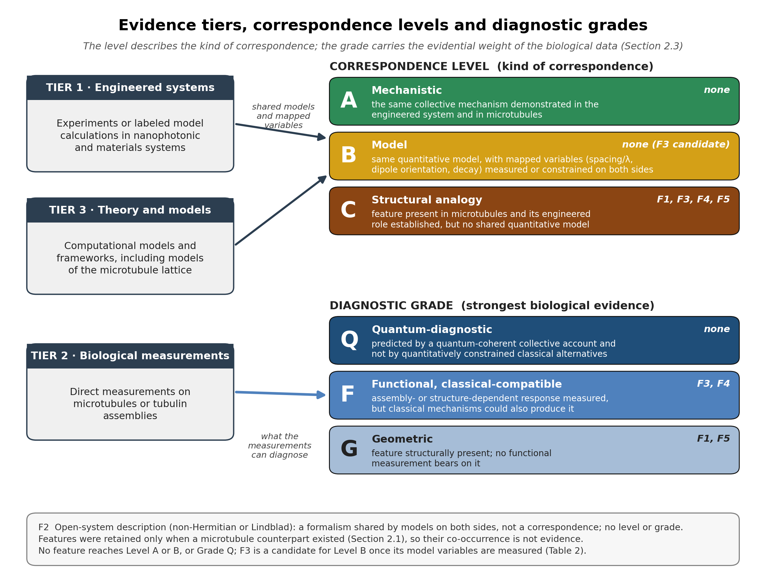
Each feature-level comparison is assigned one of three correspondence levels:
- Level A (Mechanistic correspondence): the same collective mechanism has been demonstrated in the engineered system and in microtubules.
- Level B (Model correspondence): the engineered and biological systems are described by the same quantitative model with explicitly mapped variables (for example, emitter spacing relative to wavelength, dipole orientation and collective decay rates), but the collective mode has not been demonstrated biologically. A shared formalism is not enough: the mapped variables must take measured or independently constrained values on both sides.
- Level C (Structural analogy): the structural feature is present in microtubules and its role in engineered systems is established, but no shared quantitative model maps one onto the other.
This tiered framework is designed to make the strength and limitations of each comparison transparent. These levels describe the kind of correspondence; the diagnostic grades below carry its evidential weight, and each assessment in Section 4 names the clause it satisfies. The open-system description (Feature 2) is not assigned a level, since sharing a formalism is not itself a correspondence (Section 2.2).
Separately from the correspondence level, the strongest biological evidence for each feature is given a diagnostic grade, because a measured response can depend on assembly without being specific to a quantum mechanism:
- Grade G (Geometric): the feature is structurally present; no functional measurement bears on it.
- Grade F (Functional, classical-compatible): an assembly- or structure-dependent response has been measured, but classical mechanisms (dielectric and hydration changes, scattering and reabsorption, Förster-type transport, protein quenching, conventional vibrational modes) could also produce it.
- Grade Q (Quantum-diagnostic): a measurement that a quantum-coherent collective account predicts and quantitatively constrained classical alternatives do not, such as nonclassical photon statistics or coherence signatures interpreted against classical optical bounds.
No feature currently reaches Grade Q (Section 4). Figure 1 summarizes the scheme.
2.4 Scope and Limitations of the Comparative Approach
Several methodological limitations apply to any cross-domain architectural comparison. First, geometric correspondence does not establish mechanistic equivalence: the same spatial organization may support different physics in different material contexts. Second, the five features identified here do not constitute an exhaustive list; other geometric properties (e.g., topological protection, defect tolerance) may prove relevant. Third, the biological measurements cited vary in maturity, from well-replicated structural characterizations to recent spectroscopic findings requiring independent confirmation. Where evidence quality differs, this is noted explicitly. Fourth, geometry does not fix the physical parameters that decide whether a collective mode survives: the dielectric response at the relevant frequency (anisotropic, dispersive, and different for wall-adjacent and luminal water), static site-energy disorder and its correlation between neighboring chromophores, the bath spectral density and its timescales, vibronic coupling, and the excitation or preparation condition. In engineered platforms these are designed and measured; in microtubules most are inferred from structure and simulation, or assumed. A correspondence that holds for the geometry can therefore fail quantitatively. The comparison cannot supply these values; it can identify which of them a given test must measure or constrain (Table 2 in Section 5.2; Section 5.4).
2.5 Relationship to Prior Work
This analysis complements the dimensional convergence synthesis (Echternach, 2026), which argued that the relevant 1–2 nm feature in microtubules is the tryptophan nearest-neighbor spacing (~1.3–1.8 nm, measured from PDB entries 1JFF and 3JAK), together with the hydrophobic pockets near these residues and the structured interfacial-water layers adjacent to the protein wall, in a confinement regime where quantum-relevant dielectric anomalies emerge across independent material systems; that analysis distinguishes this scale from the protofilament center-to-center spacing (~5 nm; 5.4 nm measured here, Section 3.1). The gap between adjacent protofilaments, measured directly here (Section 4.5, Figure 2), is narrower: a sub-nanometre constriction between laterally contacting protofilaments that opens into a groove. The present study turns from environmental protection (the argument that confinement shields quantum states) to architecture: which geometric variables that engineered systems show can matter for collective behavior have counterparts in the microtubule lattice, and which of those an experiment can perturb. Together, the two analyses suggest where to look and what to vary when testing for collective quantum phenomena in cytoskeletal assemblies; whether such phenomena occur depends on environmental conditions that neither analysis fixes (Section 2.4).
The present analysis is also distinct from the two 2026 reviews. Gassab and Craddock (2026) survey the evidence for quantum effects in the cytoskeleton; Bilezikian et al. (2026) survey it for ciliary proteins and articulate the shared physical features (a “quantum toolbox”) common to quantum-enabled biological systems. The contribution here is neither a survey of that evidence nor shared-features reasoning as such. The engineered platforms supply motivation: they show which geometric variables can matter for collective behavior, and so which are worth perturbing in microtubules. By the assessments in Section 4, however, none of them supplies a mechanism at microtubule parameters, and the four experiments in Section 5.3 rest on microtubule measurements and models rather than on the engineered side. The load-bearing contributions are therefore the structural measurements from PDB coordinates (Sections 3.1 and 4.5), the inventory of what the microtubule models assume against what has been measured (Table 2), and the experimental designs with their controls for classical artifacts (Section 5.3).
3. Background
3.1 Microtubule Structural Architecture
Microtubules are hollow cylindrical polymers assembled from α/β-tubulin heterodimers, with an outer diameter of approximately 25 nm and an inner lumen diameter of approximately 15 nm. Each dimer spans approximately 8 nm along the protofilament axis (Kikkawa et al., 1994), and most commonly 13 protofilaments associate laterally to form a tube (Chaaban & Brouhard, 2017); in microtubules reassembled in vitro, with 13 or 14 protofilaments, the tubulin monomers lie on a left-handed 3-start helix with an axial stagger of about 0.9 nm between adjacent protofilaments (Mandelkow et al., 1986). In the predominant B-lattice, β-tubulin subunits follow a helical path with a 12 nm pitch and an 8 nm axial repeat, which cannot close symmetrically and requires a seam of α–β lateral contacts (Kikkawa et al., 1994; Mandelkow et al., 1986), observed as a single seam in near-atomic cryo-EM reconstructions (Zhang et al., 2015). The seam is a discontinuity in the α/β lattice register; the handedness of the helix is the same on both sides of it. Adjacent protofilaments are 5.4 nm apart center to center (monomer centroids in PDB 3JAK, measured here; Data Availability Statement). The seam provides a biologically specific form of broken lattice symmetry; it is not equivalent to an engineered bilayer twist angle, but it motivates testing whether lattice-level chirality contributes measurably to polarization-resolved emission (Section 4.1). While the 13-protofilament configuration is dominant in most mammalian cells, purified tubulin assembles MTs of 9 to 16 protofilaments in vitro, and although MTs within a cell are mostly uniform in protofilament number, the number differs between cell types: in C. elegans, most neurons have 11-protofilament MTs while the six touch-receptor neurons have 15-protofilament MTs (Chalfie & Thomson, 1982; Chaaban & Brouhard, 2017), a canonical example of stable native variation. This natural variation provides experimental access to PF-number-dependent phenomena without requiring nucleotide-analog perturbation (relevant to the chirality-disruption arm of Prediction 4 in Section 5.3).
Within this lattice, tryptophan residues are geometrically organized at high density. Each α/β-tubulin dimer contains 8 tryptophan residues, and their spacings are within the near-field coupling range for UV chromophores (Babcock et al., 2024). We measured them from the deposited coordinates (PDB entries 1JFF and 3JAK; the structural papers (Löwe et al., 2001; Zhang et al., 2015) are cited only as the source of these coordinates and do not themselves discuss tryptophan positions), using the method of the companion analysis (Echternach, 2026): each indole centroid is the mean of the nine non-hydrogen ring atoms, and every pair is reported as a centroid–centroid and a closest ring-atom distance. W346 and W407 form the closest pairs at each ~4 nm tubulin boundary: αW407–βW346 at 1.53 nm within the dimer in 1JFF and 1.57 nm in the 3JAK lattice, and αW346–βW407 at 1.36 nm across the dimer boundary (closest ring atoms 1.10 and 1.17 nm within the dimer in 1JFF and 3JAK, and 0.99 nm across the boundary), creating a continuous longitudinal tryptophan chain along each protofilament. The other four tryptophans lie further from their nearest neighbor (αW388 1.8–1.9 nm, βW103 1.5–2.1 nm, αW21 and βW21 2.5–2.7 nm). In 3JAK, intra- and inter-dimer contacts were assigned from the nucleotide sites (the non-exchangeable GTP of α faces its intra-dimer β, and the exchangeable nucleotide of β faces the next α; the two criteria agree). Across adjacent protofilaments the closest tryptophans are 4.79 nm apart (4.60 nm between the nearest ring atoms), which places short-range coupling in a predominantly longitudinal geometry. All 396 measured pair distances (the 28 pairs within the 1JFF dimer, and 368 pairs in 3JAK between chains in intra-dimer, inter-dimer, lateral and diagonal contact, with no pairs counted within a chain), the input checksums and the code are in a companion notebook (Data Availability Statement). Open-system modeling points to the same pair: in a Lindblad master-equation treatment of the eight-tryptophan dimer network built on 1JFF coordinates, the αW407–βW346 pair shows the largest pairwise coherence and logarithmic negativity when the network starts in its superradiant or its most subradiant eigenstate, and βW346 appears in three of the four most coherent pairs in both cases (Gassab, Pusuluk, and Craddock, 2026, Figures 2–3); under a uniform coherent preparation the leading pairs differ. Structural geometry and the correlation model thus point to the same W346–W407 coupling, though the model uses the same 1JFF coordinates and so is not an independent test; βW103 is not prominent in this analysis, so the corroboration applies specifically to the W346–W407 link rather than to the broader tryptophan cluster. A single micron-length MT therefore contains approximately 13,000 tryptophan emitters arranged as a quasi-2D helical array. The interior lumen is occupied by structured water whose properties are sensitive to confinement geometry and whose presence is functionally required for MT electrodynamic behavior (Sahu et al., 2013). These structural features (chiral helical periodicity, extended chromophore arrays, hierarchical assembly from residue to filament to bundle, and nanoscale water confinement) are the structural counterparts examined in Section 4.
3.2 Prior Quantum Proposals and Recent Measurements
Quantum-mechanical proposals for MT function have a long history, beginning with Hameroff and Penrose’s Orch-OR theory (1996) and generating substantial debate over decoherence timescales; this debate is not the subject of the present analysis and is addressed briefly in Section 5.6. What is relevant here is that experimental measurements and computational results now bear on the view that MT geometry shapes optical behavior: Babcock et al. (2024) measured a 35% quantum yield enhancement (at 295 nm excitation) in MT assemblies relative to tubulin dimers (about 29% relative to free tryptophan); Kalra et al. (2023) documented anomalous energy migration that conventional Förster theory does not fully account for; and Craddock et al. (2017) computed that anesthetic binding shifts collective dipole oscillation modes of tubulin, with the predicted shift correlating with clinical potency. These measurements shift the question from whether MTs could support quantum phenomena in principle to whether their specific geometry is the kind of architecture that, in engineered systems, enables collective quantum phenomena, and what else such phenomena would require in a biological setting. Each is examined in detail below.
4. Results
Section 4.3 (Extended 2D Emitter Arrays) is developed in most detail because it has the most biological data. The features are presented in conceptual order, not ranked.
4.1 Helical Lattice Periodicity
Engineered evidence. Structural chirality produces handedness-dependent optical responses in engineered platforms, but every example cited here is resonant: the structures are comparable in size to the wavelength. Wang et al. (2026) reported chiral orbital lasing in a twisted bilayer metasurface with a 22.62° inter-layer twist angle, which the source attributes to helical and non-Hermitian couplings between collective guided resonances rotating clockwise and counterclockwise (Wang et al., 2026). Choi et al. (2025) fabricated 3D-printed, gold-coated helical arrays embedded in PDMS (axial pitch 250 μm, major radius 75 μm, 200 μm spacing) and measured broadband ellipticity of up to about 40° between 0.2 and 0.5 THz, which they attribute to the helices’ intrinsic chirality, multiple resonance pathways (intra- and inter-helix coupling), asymmetric interaction with circularly polarized light, and structural dispersion (Choi et al., 2025); at these frequencies the wavelength is only about one to seven times the helix pitch. Chen et al. (2026) attached individual chiral gold nanocubes (~150 nm edge) to silver nanowires and showed that, under linearly polarized 633 nm excitation, surface plasmon polaritons propagate preferentially toward one end of the nanowire according to the nanocube’s handedness, which they attribute to the nanocube’s chiroptical (plasmonic-resonance) response combined with the photonic spin-Hall effect and its plasmon coupling to the nanowire (Chen et al., 2026). These studies show that structural chirality can select optical handedness in resonant structures; none of them operates in the long-wavelength regime of the microtubule lattice.
Biological evidence. Microtubules most commonly contain 13 protofilaments, whose tubulin monomers lie on a left-handed 3-start helix; in the predominant B-lattice, β-tubulin subunits follow a 12 nm-pitch helical path that requires a seam of α–β lateral contacts (Kikkawa et al., 1994; Mandelkow et al., 1986; Section 3.1). The seam is a discontinuity in the α/β lattice register, not a change of handedness. Sahu et al. (2013) measured longitudinal four-probe conductivity and electromagnetic resonance in single MTs, and reported that these properties depend on the interior water channel (Sahu et al., 2013). That work does not measure conductance along helical paths or test chirality, so it is not offered here as functional evidence for helical periodicity; the biological evidence for this feature is structural.
Correspondence assessment (Level C). The correspondence is a structural analogy: both kinds of system are chiral, but no shared quantitative model maps one onto the other. The MT helical lattice operates at λ/a ≈ 23 for the ~280 nm tryptophan absorption band against the ~12 nm helical pitch (about 28–29 at the 330–350 nm emission maximum), far into the long-wavelength regime, whereas the engineered examples are resonant (λ/a of about 1–7 for Choi et al., 2025; a single particle about a quarter of the wavelength in size for Chen et al., 2026). No cited engineered system shows a chirality-driven optical effect at the microtubule’s ratio, so the analogy does not supply a mechanism. In the long-wavelength limit, lattice chirality could still enter through the coupling geometry of the helically arranged transition dipoles; whether this produces an emission asymmetry beyond the bulk circular dichroism of tubulin has not been measured, and Prediction 4 is designed to find out. Diagnostic grade G: the biological support is structural only.
Caveat. Bragg-type mode selection requires λ/a ≈ 1 and is not available to the MT lattice at UV wavelengths, and the resonant mechanisms of the engineered examples are not available either. A positive Prediction 4 result would show that lattice chirality affects tryptophan emission, a geometry-dependent chiroptical effect (Grade F) that coupled-dipole models can produce classically; a null result would indicate that any lattice contribution is below the bulk-CD floor or is averaged out by protein dynamics within the emission lifetime.
Testable prediction. Hypothesis: The MT helical lattice contributes a handedness-dependent asymmetry to UV fluorescence emission beyond the bulk circular dichroism of tubulin. Experiment: Polarization-resolved fluorescence spectroscopy of intact MT assemblies vs. free (unpolymerized or depolymerized) tubulin dimers, with circular dichroism (CD) of the same samples as the bulk-chirality control. Expected outcome: Intact MTs show asymmetric emission in left- vs. right-circular polarization channels with a magnitude that exceeds the linear bulk-CD prediction and falls more steeply than the bulk CD when the lattice is disrupted (a nonzero residual over the classical coupled-dipole prediction, as defined in Prediction 4). Falsification: No polarization asymmetry above the noise floor, or asymmetry fully accounted for by the linear bulk-CD scaling without a lattice-geometry contribution. This corresponds to the polarization-signature arm of Prediction 4 in Section 5.3, which also includes a multi-arm lattice-disruption design to discriminate geometric from biochemical contributions to any observed polarization signal.
4.2 Open-System Description (Shared Formalism)
Engineered evidence. Quantum optical systems coupled to environments are naturally described by non-Hermitian effective Hamiltonians, where imaginary components represent decay channels. Rather than suppressing collective behavior, controlled gain-loss asymmetry can create and stabilize it. Anisur and Choudhury (2026) modeled the open Dicke model (two-level atoms in a single-mode cavity with cavity decay, written as a Lindblad master equation) under a quasi-periodic Fibonacci drive, and found time-quasicrystal order in the thermodynamic limit (semiclassical analysis) and, in exact-diagonalization calculations for two to five qubits, a finite lifetime τ* that grows linearly with qubit number, which the authors extrapolate to stability in the thermodynamic limit (Anisur & Choudhury, 2026). The quasi-periodicity here is in time, in the drive sequence, not in the spatial arrangement of the emitters. This result, from a preprint (Tier 3 evidence), indicates, in the words of the authors’ introduction, that “the interplay of dissipation and quasi-periodic driving provides a powerful route to realize stable non-equilibrium phases of matter” (Anisur & Choudhury, 2026, p. 1). Bychek et al. (2025) predicted mirrorless superradiant lasing in a dense emitter chain in which unpumped atoms, acting as narrow-band out-coupling antennas through resonant dipole-dipole interaction, enhance emission power and spectral purity (Bychek et al., 2025). The source frames the unpumped emitters as an effective high-Q resonator that filters and amplifies the broadband gain of the pumped fraction; it does not use non-Hermitian gain–loss language, and placing it here is our interpretation. This theoretical work suggests that environmental coupling can be a resource rather than a nuisance when managed through appropriate geometric arrangement.
Biological evidence. Babcock et al. (2024) modeled the tryptophan network of MT lattices using a non-Hermitian Hamiltonian of the form H_j = E_j − iΓ_j/2, where E_j is the site energy and Γ_j the radiative decay rate (Babcock et al., 2024). This formalism (of the same family as the Lindblad treatment used by Anisur et al.) captures the open-system character of biological chromophore networks embedded in a dissipative protein environment. Collective superradiant states emerge from this model with enhanced quantum yield relative to the single-emitter baseline. The effective non-Hermitian Hamiltonian is, however, an approximate description: it does not preserve the trace of the density operator once radiative decay is included, and is properly understood as the no-jump limit of a more complete treatment. Gassab, Pusuluk, and Craddock (2026) supply that treatment for small networks: they propagate a trace-preserving Lindblad master equation, built from the same coupling and radiative-decay matrices, for a single tubulin dimer (eight tryptophans) and for embeddings of up to two spirals. For larger assemblies (up to 100 spirals) they note that direct Lindblad simulation is not practical and return to the effective non-Hermitian Hamiltonian to obtain eigenmodes and radiative decay rates. The biological side therefore rests on a Lindblad treatment for small networks and on the approximate non-Hermitian model at lattice scale.
Their analysis resolves the collective dynamics into two channels: a superradiant preparation whose pairwise coherence drops sharply within about 1 ns and is largely extinguished within 5 ns, and a subradiant preparation whose population and coherence remain elevated across the 0–80 ns window shown, with entanglement decaying over roughly the first 40 ns, in the eight-site case. The longer-lived retention channel is the more plausibly functional one. In the same paper’s non-Hermitian eigenmode analysis of ordered lattices, from a single dimer to 100 spirals, increasing size shortens the most superradiant lifetime and lengthens the most subradiant radiative lifetime into the millisecond range (the cooperative suppression of the dark-state decay width also reported by Celardo et al., 2019, whose superradiant decay width grows in proportion to emitter number). This is a radiative lifetime, the time a dark state sits without emitting, not a coherence time, because the model dissipates through radiative channels only and does not bound the non-radiative dephasing that sets the functional coherence limit in a warm, aqueous environment. The retained nonclassical correlations within the model are therefore the tens-of-nanosecond entanglement, not the much longer radiative lifetime. Kalra et al. (2023) provided complementary experimental evidence: exciton diffusion lengths of 6.6 nm in microtubule lattices that conventional Förster theory (accommodating tryptophan–tyrosine interactions) “does not sufficiently explain” (Kalra et al., 2023, abstract). Their FRET-based kinetic Monte Carlo model reproduces hopping dynamics but underestimates the observed transport range, consistent with environment-mediated transport enhancement of the kind described by non-Hermitian models.
Status of this dimension (no level assigned). Both sides are modeled with open-system methods: the engineered model of Anisur and Choudhury (2026) is a Lindblad master equation, and on the biological side the same trace-preserving formalism has been applied to networks of up to two spirals (Gassab, Pusuluk, and Craddock, 2026), while lattice-scale results come from an effective non-Hermitian Hamiltonian (Babcock et al., 2024; Gassab, Pusuluk, and Craddock, 2026). Sharing a formalism is not a correspondence (Section 2.2), and the physics differs: the engineered model is a driven Dicke system in a cavity whose result is time-quasicrystal order, whereas the microtubule models describe undriven radiative relaxation, with transient preservation and redistribution of correlations and no cavity or bath beyond radiative loss. What the shared description offers is a common set of variables (emitter positions and orientations, coherent couplings and collective decay rates) that the experiments of Section 5.3 can constrain. No measurement yet probes a signature specific to the non-Hermitian description.
Caveat. The biological evidence rests primarily on a theoretical model (Babcock et al.) supported by indirect experimental measurements (Kalra et al.). No direct measurement of superradiant burst dynamics in MT tryptophan networks has been reported. The Anisur et al. preprint has not yet undergone peer review and should be weighted accordingly.
Testable prediction. Hypothesis: If non-Hermitian dynamics are operative in MT tryptophan networks, coupling between adjacent MTs in a bundle should alter collective decay rates in a way that is inconsistent with independent emitter behavior. Experiment: Time-resolved UV fluorescence of MT bundles vs. isolated MTs of matched total length, measuring fluorescence decay kinetics. Expected outcome: MT bundles should show superlinear scaling of early-time emission intensity (superradiant burst) with MT number, and altered decay timescales relative to isolated MTs. Falsification: Decay rates scale linearly with emitter number (classical additive behavior), indicating no inter-tubule coupling of collective modes. Controls: as for Prediction 1 (Section 5.3): matched optical density, inner-filter and scattering corrections, and the bundling agent alone; a superlinear result that survives them is necessary but not sufficient for inter-tubule collective coupling.
4.3 Extended 2D Emitter Arrays
Engineered evidence. Spatial organization of quantum emitters can change their cooperative radiative response, but the effect depends not on array geometry alone: transition wavelengths and dipole orientations, emitter-to-emitter detuning, local electromagnetic density of states, coupling to photonic modes, preparation, and radiative and nonradiative losses all enter. Eyles et al. (2026) theoretically proposed Dicke-like superradiance for extended two-dimensional arrays of emitters coupled through a metasurface bound state in the continuum (BIC). Their model predicts that efficient coupling to a nonlocal, high-Q BIC mode, quantified through a coupling-efficiency parameter, can mediate collective emission even when emitter separations exceed the free-space wavelength. This is a useful demonstration within a model, not an experimental demonstration, and the existence of a comparable BIC mode in microtubules has not been established. Bychek et al. (2025) likewise theoretically predicted mirrorless superradiant lasing from a densely spaced one-dimensional emitter chain containing partially pumped and unpumped atoms. Its cavity-free result demonstrates that a physical optical cavity need not be included in that model, but the pumped, driven steady-state setting differs fundamentally from native, undriven tryptophan fluorescence. Its chain is also short: eight emitters, with the collective enhancement appreciable only at spacings below about a quarter of the transition wavelength, so the whole chain spans less than two wavelengths. For large arrays in free space, rigorous bounds on the maximal collective decay rate rule out superradiant lasing in one-dimensional arrays, because the emitted intensity never scales superlinearly with emitter number, while leaving it possible in two- and three-dimensional arrays (Mok et al., 2026). The cavity-free result therefore applies to short, subwavelength-spaced chains and does not carry over to the length of a single microtubule, which, at about 25 nm across against a 280 nm transition wavelength, we treat as a quasi-one-dimensional array. These engineered models motivate geometric and photonic variables to perturb; they are not evidence that an analogous collective mode operates in a microtubule. Rodríguez-Rubio et al. (2025) provide a complementary molecular example of architecture-dependent electronic behavior: experimentally observed aromatic and antiaromatic ring currents in covalently conjugated porphyrin nanobelts. That result concerns global π-conjugation and ring currents, not cooperative UV emission from distinct aromatic residues, and should be understood as an analogy in geometric control of electronic structure rather than as an emitter-array benchmark.
Microtubule architecture and measurements. The α/β-tubulin dimer carries eight tryptophan chromophores organized into a repeating cylindrical lattice. This gives microtubules a dense, anisotropic network of potential optical and excitation-transfer sites, whose interactions depend on distances, relative orientations, disorder, and their heterogeneous protein/water environment. Babcock et al. (2024) measured assembly-dependent tryptophan fluorescence quantum yields (QYs). Their reported values at 295-nm excitation were 14.7 ± 1.6% for microtubules and 10.9 ± 1.3% for tubulin dimers, a roughly 35% relative increase; the corresponding 280-nm tryptophan-attributed values were 17.6 ± 2.1% and 10.6 ± 0.6%, a roughly 66% relative increase. The same study measured free tryptophan at 11.4 ± 1.1% (295 nm) and 12.4 ± 1.1% (280 nm): the dimer value lies below free tryptophan, and against free tryptophan the microtubule excess is about 29% (295 nm) and 42% (280 nm), so part of the dimer-to-microtubule difference may be relief of quenching present in the dimer rather than enhancement over an uncoupled chromophore. The study also modeled radiative cooperative modes in ordered tryptophan geometries. Its measurements establish an optical difference between preparations of assembled and unassembled tubulin, but steady-state QY alone cannot establish an increased collective radiative rate: changes in nonradiative decay, hydration, absorption normalization, morphology-dependent scattering or collection, and reabsorption may contribute. The original paper notes a scattering contribution to microtubule absorption that was corrected and that the relevant joint lifetime/radiative-rate measurement had not been performed.
Kalra et al. (2023) independently inferred a two-dimensional photoexcitation diffusion length of 6.6 ± 0.1 nm from tryptophan fluorescence-lifetime quenching measurements in microtubules. This exceeded their conventional Förster-model estimates of 1.11 nm for the intertyrosine channel and 1.54 nm when tyrosine–tryptophan interactions were included. Diffusion was sensitive to assembly state and reduced in the presence of etomidate and isoflurane; changing the average protofilament number from 13 to 14 did not significantly alter the estimated length. These results make optical migration a distinct and informative endpoint from QY. They do not by themselves identify a delocalized coherent carrier or exclude more complete classical hopping, heterogeneous dielectric, and vibronically coupled transport models. In particular, the null result for 13- versus 14-protofilament lattices requires any proposed helicity-sensitive mechanism to make predictions more specific than a generic ‘more ordered geometry means more transport’ expectation.
Additional spectroscopy and modeling: distinct evidence categories. Zhang et al. (2022) reported assembly- and hydration-sensitive Raman spectra, with multiple lines that they interpreted as possible Fano-like resonances. The study found candidate asymmetrical profiles in both aqueous tubulin and aqueous microtubules, and discussed alternative assignments for some bands, notably features near the tyrosine doublet. These measurements motivate controlled spectroscopy of environmental and assembly dependence; they do not establish that a Fano mode appears uniquely on polymerization or directly reveal sustained inter-residue quantum coherence. Craddock et al. (2017) studied anesthetic effects by computational docking, molecular polarizability calculations, and normal-mode analysis of London-induced dipole oscillators. Their reported high correlation (R² = 0.999) relates a predicted anesthetic-induced mode-frequency shift to known anesthetic potency; it is not an experimental measurement of suppressed microtubule THz oscillations. Furthermore, these induced-dipole modes should not be conflated with optical Frenkel-exciton exchange between tryptophans. Modeling by Shirmovsky and Chizhov (2023) predicts conditional excitation and entanglement transfer in a dipole-coupled tryptophan chain, but such model results do not provide an independent biological observation of entanglement. Taken together, these sources identify experimentally accessible endpoints and theoretical possibilities; they do not all independently measure the same physical mechanism.
Classical alternatives and discriminating conditions. The most immediate explanation of assembly-dependent QY is altered local tryptophan photophysics, especially solvent exposure, conformational rigidity, and nonradiative decay. However, a rigorous null cannot stop with independent-emitter quenching. Assembly also changes the optical density, angular scattering, anisotropic refractive-index environment, absorption and reabsorption pathways, and distribution of local transition frequencies and dipole orientations. Classical multi-step Förster transport and electrodynamic coupling can themselves produce geometry-dependent and sometimes nonlinear signatures. A lattice-dependent signal therefore establishes a geometric effect only after optical-transport and chemical confounds have been bounded; it does not automatically establish a quantum-coherent collective state.
The useful distinction is among three propositions: (i) a molecular lattice presents a coupled emitter geometry; (ii) assembly changes an experimentally measured optical response; and (iii) a particular nonclassical cooperative mechanism is required to explain that response. Current measurements strongly support the first two statements, while the third remains open. In particular, a structurally connected tryptophan network does not imply an electronically delocalized state robust to site-energy detuning, environmental fluctuations or decoherence. An adequate quantitative test must specify the optical-electronic Hamiltonian, the biological dielectric response at the relevant frequency, excitation/preparation conditions, and both radiative and nonradiative bath coupling. Small geometry or dielectric changes may affect observables, but their magnitude and sign must be calculated or measured rather than assumed.
Correspondence assessment (Level C). Extended, organized chromophore networks exist in the microtubule lattice, and experimental evidence shows assembly-conditional fluorescence and excitation-migration behavior. Engineered nanophotonic theory demonstrates that cooperative radiative responses can depend sharply on emitter topology and coupling to optical modes. However, the cited engineered studies do not experimentally establish that microtubules realize the same mode structure; the key biological measurements have not yet distinguished a specific quantum-cooperative mechanism from the full relevant family of classical alternatives. The free-space collective-emission models applied to engineered emitter chains (Bychek et al., 2025) and to the tryptophan lattice (Celardo et al., 2019; Babcock et al., 2024) share the dipole–dipole coupling and collective-decay description, with emitter spacing relative to wavelength, dipole orientation and collective decay rates as the variables to be mapped; the BIC-mediated model of Eyles et al. (2026) adds a photonic mode with no microtubule counterpart. That description is, however, generic to any set of near-field-coupled chromophores, and on the microtubule side its variables are not yet constrained: the transition dipole differs by a factor of about three between models, screening is omitted or assumed, and disorder and bath coupling are assumed or absent (Table 2). We therefore assign Level C (structural analogy), with a candidate model correspondence that would reach Level B once those variables are measured or independently constrained. The value of the analogy is that it specifies the independent variables, competing mechanisms and measurements needed to move from architectural resemblance to mechanistic identification. Diagnostic grade F: the quantum yield enhancement and the 6.6 nm migration length are measured assembly-dependent responses that the classical alternatives above could also produce.
Testable predictions and controls. The clearest next step is a multi-observable study of microtubule fragments of controlled length, isolated filaments and bundles. Samples should be matched for tryptophan count, polymerization and nucleotide/stabilizer chemistry, concentration, excitation conditions, hydration, temperature, optical depth and measured morphology. Absolute fluorescence QY should be paired with time-resolved fluorescence decay, absorbance/extinction, integrating-sphere or angle-resolved scattering measurements, emission anisotropy, and explicit reabsorption/inner-filter corrections. Quencher-dependent diffusion measurements can test whether structural perturbations change transport separately from QY. A superlinear size or bundle dependence would be evidence of a nonadditive architecture-dependent response; it would not by itself reject classical collective or optical-transport mechanisms. Support for a quantum-cooperative explanation would require agreement with a specified collective dynamics model, exclusion of quantitatively constrained classical alternatives, and where feasible a direct coherence or nonclassical photon-statistics diagnostic interpreted against classical optical bounds. Anesthetic titrations and chirality perturbations can supply orthogonal tests only if protein stability, binding, scattering and local solvent conditions are measured in parallel. Failure of such experiments to identify a nonclassical contribution would narrow the mechanism without negating the observed assembly-dependent optical biology.
4.4 Hierarchical Nesting
Engineered evidence. Hierarchically nested structures (smaller-scale periodic motifs embedded within larger-scale frameworks) are proposed to produce collective behavior that neither scale shows alone, but the engineered support cited here is weak. Singh et al. (2026) reported room-temperature soliton–polariton condensation in a self-assembled hierarchical helical-nanowire gel, with partial coherence (g⁽²⁾(0) ≈ 1.26), and attribute nearly two orders of magnitude of photon confinement to nested interference and spin–momentum locking across fractal layers (Singh et al., 2026); no single-scale control is reported, and the work comes from the same group as two of the biological sources below. Guo et al. (2025) reported coherent normal-state charge transport in microstructures of the kagome metal CsV₃Sb₅ below about 30 K, seen as magnetoresistance oscillations from interlayer Aharonov–Bohm interference that persist beyond the single-particle mean free path and switch at angles set by the shape of the sample cross-section, which the authors attribute to a long-range coherent many-body state sensitive to the geometry of micron-scale confinement (Guo et al., 2025). This shows that geometry can shape collective electronic coherence at cryogenic temperature; it is not evidence of hierarchical nesting or of mode redistribution across scales. Bilezikian et al. (2026) list quasicrystalline and topological order, as approximated in cytoskeletal architectures and their hydration water, as an outstanding question for the field (their Table 2); the dissipative Dicke time quasicrystal (Anisur and Choudhury, 2026) bears on it only loosely, because its quasi-periodicity lies in the drive sequence rather than in spatial geometry.
Biological evidence. The MT cytoskeleton exhibits hierarchical nesting across at least five spatial scales: individual tryptophan residues (~0.3 nm) → tubulin dimers (~8 nm) → protofilaments (linear chains of dimers, spaced 5.4 nm center to center in the wall; Section 3.1) → microtubules (~25 nm diameter, microns long) → MT bundles and centrioles (hundreds of nm). The protofilament level adds longitudinal continuity of the dimer chain, and the microtubule level adds lateral closure of 13 protofilaments into a tube. Babcock et al. (2024) measured quantum yields for two assembly levels only, microtubules and tubulin dimers (14.7 ± 1.6% versus 10.9 ± 1.3% at 295 nm excitation; Section 4.3); larger assemblies could not be measured because of scattered light and sample purity problems (Babcock et al., 2024). The architecture-by-architecture quantum yield increase across the hierarchy in that paper comes from its theoretical model, not from measurement. The measured dimer-to-MT difference is consistent with, but does not demonstrate, a collective contribution arising at the assembly level.
Cantero et al. (2018) recorded spontaneous, self-sustained electrical oscillations in loose-patch-clamped rat brain MT bundles, with a prominent 39 Hz fundamental frequency and amplitude envelopes that the authors describe as fractal; they describe the bundle response as similar to, but richer than, the response they had earlier reported for 2D MT sheets. Taxol (10 μM) inhibited the oscillations, which the authors used to confirm that the activity is intrinsic to the MT bundle, and they state that the data should be considered qualitatively (Cantero et al., 2018). The study did not compare assembly levels directly. It is consistent with assembly-dependent electrical behavior, from a group independent of the others cited here, but does not show that hierarchical organization redistributes collective modes.
Saxena et al. (2020) reported a self-similar ‘triplet of triplet’ pattern of electromagnetic resonance bands in a single tubulin protein (bands from MHz to THz), a single microtubule (kHz to GHz) and the axon initial segment of a neuron (Hz to MHz). Hameroff et al. (2026), a group that includes Bandyopadhyay, interpret these resonance data as showing that microtubules behave as ‘fractal time crystals’ and link this to consciousness; the interpretation is speculative and has not been independently replicated.
Theoretical modeling bears on assembly dependence on the optical side. Celardo et al. (2019) showed that the native tryptophan lattice supports a superradiant lowest exciton state that emerges, via supertransfer coupling between smaller blocks, once an ordered segment exceeds ~12 spirals (~108 nm). Gassab, Pusuluk, and Craddock (2026) extended this with a Lindbladian treatment showing that embedding a tubulin unit into progressively larger assemblies (single tubulin → two tubulins → one spiral → two spirals) changes which site pairs carry the most coherence, lowers coherence amplitudes within the focal tubulin and adds oscillatory exchange with its neighbors; in a separate non-Hermitian eigenmode analysis of ordered lattices of up to 100 spirals, where they judge Lindblad simulation impractical, superradiant and subradiant radiative lifetimes diverge with size. These models share authors, coordinates and a radiative-loss-only treatment (Section 2.1), so they are one modeling line rather than independent confirmations.
Correspondence assessment (Level C). Hierarchical assembly is structurally present in microtubules (Section 3.1), but no shared quantitative model maps an engineered nested structure onto microtubule assembly levels, and the engineered support is thin: Singh et al. (2026) is a single-group report with partial coherence, and Guo et al. (2025) concerns geometry-sensitive electronic coherence, not nesting. The biological measurements (Babcock et al., 2024, optical, dimer versus MT; Cantero et al., 2018, electrical, qualitative; Saxena et al., 2020, electromagnetic resonance bands) show assembly- or scale-dependent behavior, but none tests whether nested organization reshapes collective optical mode structure, which is the decisive experiment. We therefore assign Level C (structural analogy). Diagnostic grade F: assembly-dependent responses are measured, but each is classical-compatible, and the Saxena and Cantero results await independent replication.
Caveat. Three Feature-4 sources share an author: Singh et al. (2026) on the engineered side, and Saxena et al. (2020) and Hameroff et al. (2026) on the biological side, all include Bandyopadhyay, and are therefore weighted as one line of work. The self-similar resonance pattern reported by Saxena et al. (2020), a journal article, has not been independently replicated, and the fractal time crystal interpretation (Hameroff et al., 2026) is speculative. The Cantero et al. measurements are reported as qualitative under loose-patch configuration by the authors themselves; they should be weighted as converging rather than quantitative evidence. Feature 4 therefore lacks an independent engineered anchor.
Testable prediction. Hypothesis: If hierarchical nesting reshapes collective mode structure in MTs, intact bundles should show qualitatively different collective optical signatures than isolated MTs containing an equal total number of tryptophan emitters. Experiment: Polarization-resolved UV fluorescence of (a) MT bundles of defined size and (b) isolated MTs with matched tryptophan count, comparing emission enhancement, decay kinetics, and polarization signatures. Expected outcome: Bundles should show collective signatures not predictable by linear superposition of isolated MT signals, specifically additional enhancement or altered decay timescales attributable to inter-tubule geometric coupling. Falsification: Bundle signatures are quantitatively predictable from linear superposition of isolated MT signals, indicating no cross-scale mode redistribution. Controls: as for Prediction 1 (Section 5.3), since bundling also changes optical density, scattering and local chemistry. In Section 5.3 this test is carried by Prediction 1 with a polarization channel added, and Prediction 1’s feasibility limits apply to it.
4.5 Nanoscale Confinement
Engineered evidence. Confinement of water and other dielectric media to nanoscale dimensions alters their electronic and dielectric properties in ways that can protect or enable quantum coherence effects. Water confined between atomically flat surfaces is dielectrically anomalous and strongly anisotropic: perpendicular to the walls, the near-surface layers become essentially non-polarizable, with a dielectric constant of about 2 (Fumagalli et al., 2018), whereas along the walls water 1–2 nm thick reaches ferroelectric-like values of about 1,000, against about 80 in bulk (R. Wang et al., 2025). Both were measured between hBN or graphite walls, and the values probably depend on surface chemistry (R. Wang et al., 2025). Reiter et al. (2016) showed that water confined within carbon nanotubes exhibits quantum coherence effects distinct from bulk water, including proton delocalization and non-classical momentum distributions (Reiter et al., 2016). These results establish that nanoscale geometric confinement is sufficient to produce qualitatively distinct water properties relevant to quantum phenomena.
Biological evidence. The MT lumen itself is approximately 15 nm in diameter and should not be equated with the 1–2 nm confinement gaps studied in engineered planar systems. The relevant biological counterparts, following the companion analysis (Echternach, 2026), are the ~1.3–1.8 nm tryptophan nearest-neighbor spacing, the hydrophobic pockets near these residues, and the structured interfacial-water layers adjacent to the protein wall; these provide local nanoscale confinement, while the larger lumen may serve as a broader aqueous cavity supporting longer-range electrodynamic coupling. Sahu et al. (2013) reported that the electronic and resonance properties of a single MT depend on the interior water channel and disappear when the atomic water core is released (Sahu et al., 2013). This water-dependence suggests that the interior water is functionally integral to the MT’s quantum-relevant properties, not a passive solvent.
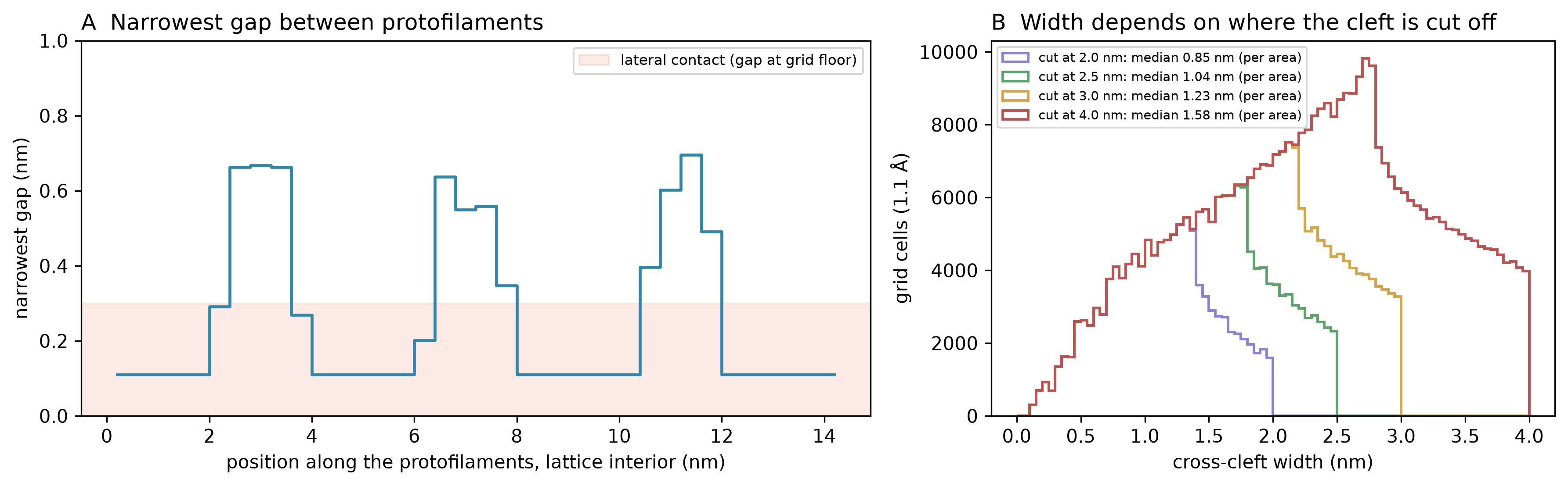
The gap between adjacent protofilaments was measured directly (Figure 2). In the 3JAK lattice (protofilaments formed by chains H+A+B+K and E+F+G+J, with EB3 and the nucleotides excluded; Bondi van der Waals radii, hydrogens excluded, 1.1 Å grid), the cross-cleft width at each empty grid point (outside every atom’s van der Waals sphere; no water-sized probe is applied) is the surface-to-surface distance across to the two protofilaments. These are gaps between van der Waals surfaces, not water-accessible space: a water molecule, about 0.28 nm across, fits only where the gap is wider than that. The protofilaments are in lateral contact along most of their length: in 4 Å slices through the interior of the lattice segment, the narrowest gap between them is at the grid floor (about 0.1 nm) in about half of the slices and never exceeds 0.70 nm (90th percentile 0.65 nm), opening once per ~4 nm tubulin repeat (Figure 2A). These values do not depend on the grid or the slicing: at grid spacings from 0.55 to 1.5 Å the maximum is 0.68–0.71 nm, the 90th percentile 0.61–0.65 nm and 56–61% of slices sit at the grid floor, and slices of 2 to 8 Å give a maximum of 0.66–0.70 nm and a dominant period of 4.0–4.8 nm; trimming only 1 nm, rather than 2 or 3 nm, from the segment ends admits end slices up to 0.91 nm. We did not repeat the analysis on a second microtubule structure. Away from these constrictions the cleft widens continuously toward the lumen and the outer surface, so it has no natural outer boundary: the width distribution of the empty space between the protofilaments follows wherever it is cut off, with a median per unit of cleft area of 0.85, 1.04, 1.23 and 1.58 nm for cutoffs at 2.0, 2.5, 3.0 and 4.0 nm (Figure 2B). A fixed width cutoff therefore sets, rather than measures, the upper end of any reported cleft range. The confinement geometry the lattice itself defines is a sub-nanometre constriction between contacting protofilaments, opening into a groove whose width depends on how far from the wall it is measured; where the protofilaments touch there is no room for water, at the widest constrictions (about 0.7 nm) there is room for roughly two water layers, and the water-accessible confinement is the groove they open into. This geometry is distinct from both the 5.4 nm protofilament center-to-center spacing and the 15 nm lumen. Fourteen of the 32 tryptophans on the two flanking protofilaments lie within 1.5 nm of the empty space between them (the notebook prints 14 of 48 because it counts all twelve loaded chains; the 14 it lists all lie on the eight flanking chains); we make no claim about their placement relative to the constrictions, and treat the tryptophan chain, which runs longitudinally within each protofilament (Section 3.1), and the inter-protofilament gap as separate features.
Correspondence assessment (Level C). The geometric counterparts of quantum-relevant confinement (tryptophan-scale spacing, a sub-nanometre constriction between protofilaments that opens into a groove, structured interfacial water) are present in MTs, and interior water has been reported to be functionally relevant to MT conductivity and resonance (Sahu et al.). The environmental conditions enabling quantum coherence in nanophotonic analogs, specifically nanoscale water confinement, are treated in detail in a companion analysis (Echternach, 2026); here we note only that the tryptophan-scale spacings and wall-adjacent water layers are the features identified there. We assign Level C (structural analogy): the confinement geometry is present, but no shared quantitative model maps confined water at hBN or graphite walls onto the protein-lined cleft, and detailed treatment is deferred to the companion paper. Diagnostic grade G: the confinement geometry is measured; the water dependence of MT conductance (Sahu et al., 2013) is an electrical response that does not bear directly on a quantum mechanism, so it is context rather than functional evidence for this feature.
Caveat. The proposed counterpart regions (tryptophan-scale spacings, hydrophobic pockets, structured wall-adjacent water) involve protein-embedded and curved-wall geometries that differ from the planar surface confinement of the Wang et al. system, and no direct measurement of MT interfacial water properties analogous to the Wang et al. or Reiter et al. systems has yet been reported. The cleft widths in Figure 2 are static heavy-atom distances from a single cryo-EM structure; hydrogens, bound water and thermal motion would change the effective width.
For a full treatment of nanoscale confinement as an enabler of quantum coherence, including dimensional convergence analysis across biological and engineered systems, see Echternach (2026).
5. Discussion
5.1 Synthesis of Correspondences
The five dimensions examined in Section 4 are not independent variables; they are structurally coupled. Helical lattice periodicity (Feature 1) constrains the geometry of the tryptophan array (Feature 3): the 3-start helical lattice positions emitters in a quasi-2D arrangement whose spacing and orientation are determined by the protofilament pitch. The open-system description (Feature 2) is not a separate structure: it is how the emitter network of Feature 3 is modeled. Hierarchical nesting (Feature 4) encompasses all the lower features, since each assembly level (dimer, protofilament, MT, bundle) inherits the geometry of the level below while adding inter-level coupling. Nanoscale confinement (Feature 5) provides the dielectric environment within which the structural features operate. This coupling means the dimensions should not be counted as independent analogies, and because they were retained only when a microtubule counterpart existed (Section 2.1), their co-occurrence is not offered as evidence. What the coupling supplies is a set of variables that the experiments of Section 5.3 can perturb together or separately.
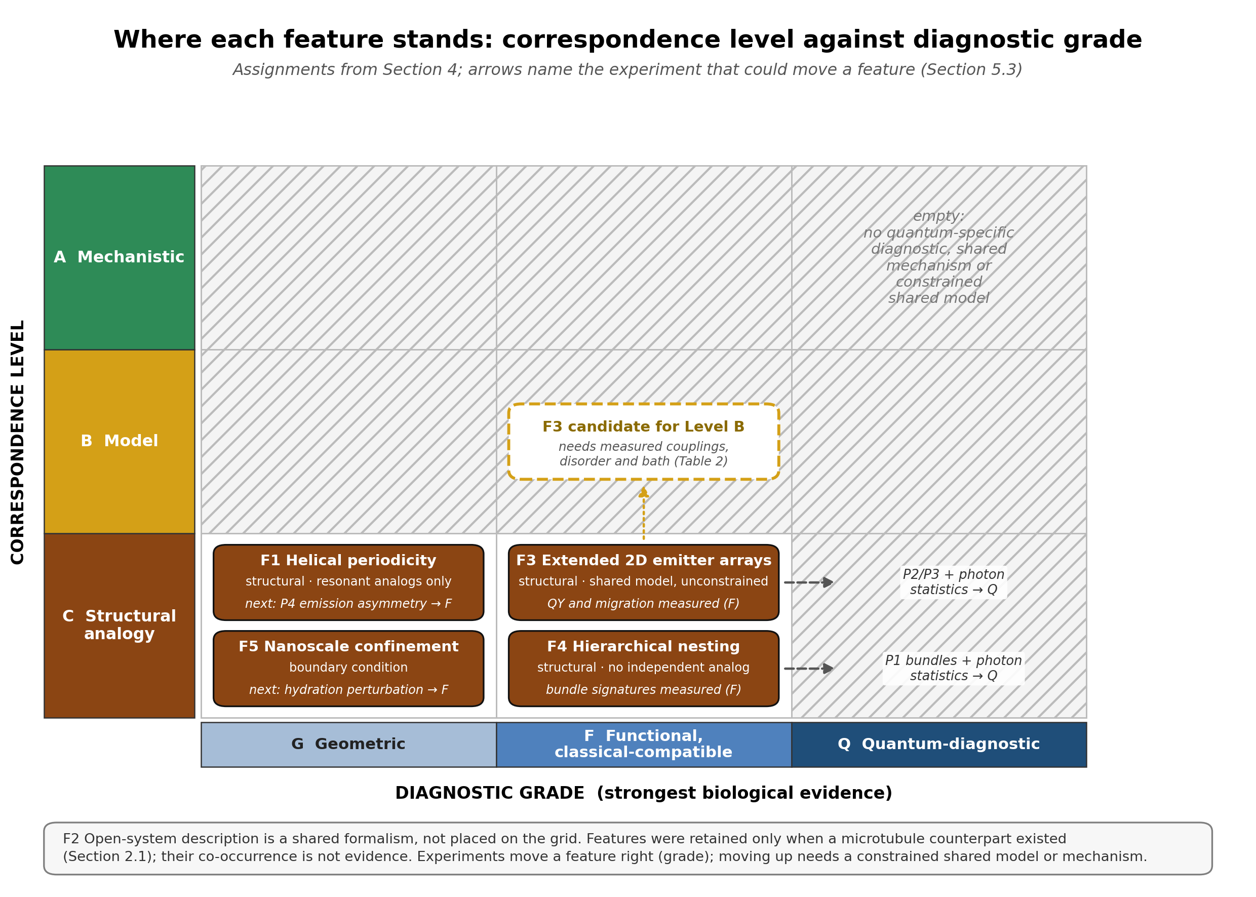
Table 1 summarizes the evidence for each feature and its correspondence level, and Figure 3 places each feature by level and grade.
| Feature | Engineered Evidence | MT Evidence | Level | Major Caveat | Decisive Experiment |
|---|---|---|---|---|---|
| Helical lattice periodicity | Wang et al. (2026): chiral orbital lasing in a resonant twisted-bilayer metasurface; Choi et al. (2025): THz ellipticity in resonant gold helices; Chen et al. (2026): handedness-dependent plasmon routing at 633 nm | Mandelkow et al. (1986); Kikkawa et al. (1994): left-handed 3-start helix, 12 nm pitch, lattice seam; Sahu et al. (2013): water-dependent longitudinal conductivity (no helical-path measurement) | C | Engineered examples are resonant (λ/a ≈ 1–7); at λ/a ≈ 23 the MT lattice has no engineered mechanistic anchor | Polarization-resolved UV fluorescence: intact MTs vs. depolymerized tubulin, with CD as bulk-chirality control |
| Open-system description (shared formalism) | Anisur and Choudhury (2026, preprint): time-quasicrystal lifetime τ* grows with system size (theory); Bychek et al. (2025): unpumped atoms enhance lasing (theory) | Babcock et al. (2024): H_eff = E_j − iΓ_j/2; Gassab, Pusuluk, and Craddock (2026): Lindblad treatment up to two spirals; Kalra et al. (2023): exciton diffusion 6.6 nm, conventional Förster insufficient | — | A shared formalism, not a correspondence; the physics differs (driven cavity vs. undriven radiative relaxation) | Time-resolved fluorescence: bundles vs. isolated MTs |
| Extended 2D emitter arrays | Eyles et al. (2026, preprint): BIC-mediated Dicke superradiance (theory); Bychek et al. (2025): cavity-free superradiant lasing (theory) | Babcock et al. (2024): 35% QY enhancement over tubulin dimers, ~29% over free Trp (295 nm), 13,000 Trp/μm-MT; Kalra et al. (2023): 6.6 nm diffusion (Förster insufficient); Craddock et al. (2017): computed R²=0.999 (theory); Zhang et al. (2022): asymmetric (Fano-like) Raman lines in both tubulin and MTs | C (candidate B) | Shared description is generic; mapped variables unconstrained (Table 2); Eyles is preprint | Anesthetic titration + array-size dependence in MT fragments |
| Hierarchical nesting | Singh et al. (2026): partial-coherence polariton condensation in a hierarchical gel (same group as Saxena); Guo et al. (2025): geometry-sensitive coherent interlayer transport in a kagome metal (≤30 K) | Babcock et al. (2024): QY dimer vs. MT (measured), intermediate levels modeled; Cantero et al. (2018): bundle electrical oscillations, 39 Hz (qualitative, loose patch); Saxena et al. (2020): self-similar EM resonance bands (tubulin, single MT, neuron) | C | No independent engineered anchor; optical mode-redistribution test outstanding | Prediction 1 with a polarization channel (bundles vs. isolated MTs, matched emitter count) |
| Nanoscale confinement | Fumagalli et al. (2018) and R. Wang et al. (2025): anisotropic dielectric at 1–2 nm (ε⊥ ≈ 2, ε∥ ≈ 1,000); Reiter et al. (2016): quantum water in CNTs | Sahu et al. (2013): water-dependent conductance; inter-protofilament gap ≤0.7 nm at its narrowest, opening into a groove (3JAK, this work) | C | Static cryo-EM geometry; curved, protein-lined cleft differs from planar confinement; the 15 nm lumen is not the counterpart | Direct mapping of MT interfacial and luminal-water dielectric response, separating wall-adjacent and lumen-core contributions. |
5.2 From Analogy to Mechanism
Geometric correspondence identifies candidate conditions, not sufficient ones. The argument presented here is that microtubule architecture contains candidate structural motifs of the kind that nanophotonic engineering associates with collective quantum phenomena. Containing such motifs does not show that the quantitative conditions the engineered systems also require (mode quality, coupling efficiency, detuning, disorder and preparation; Table 2) are met. This distinction matters, and we have tried to enforce it consistently throughout.
The structural correspondences identified here show that some of the spatial conditions associated with collective phenomena in engineered systems are present in microtubules. The ~1.4–1.6 nm spacing along the W346–W407 tryptophan chain is deeply subwavelength (d/λ ≈ 0.005 at 280 nm); whether it supports collective behavior depends not on the spacing alone but on how the computed coupling (~50–60 cm⁻¹; Celardo et al., 2019; Babcock et al., 2024) compares with site-energy disorder (estimated at 50–300 cm⁻¹) and with bath coupling (a Stokes shift more than thirty times larger), none of which is measured in microtubules (Table 2). The 13-protofilament helical lattice is chiral, although at a wavelength-to-pitch ratio far beyond that of the resonant engineered chiral structures. These are correspondences in structural parameters that influence collective behavior in engineered systems, not merely visual analogies, but they are not shared mechanisms.
These correspondences do not, however, establish that the same physical mechanism is operative. No photonic cavity is present; the protein matrix is an open, lossy environment. Room-temperature operation in an aqueous, dynamically disordered medium introduces phonon bath properties, spectral densities, and reorganization energies that differ from those of semiconductor quantum dots or atomic emitters. Geometry is not mechanism, but it is often the condition under which mechanism becomes experimentally realizable. Whether it does so in microtubules turns on the environmental parameters listed in Section 2.4, and uncertainty in those parameters cuts both ways: it leaves room for a collective mechanism that current models miss, but it is not evidence that one operates. Where multiple measurements and theoretical frameworks bear on the same question (Section 4.3 sets out the measurements and the classical alternatives for Feature 3, and concludes that the key biological measurements have not yet distinguished a quantum-cooperative mechanism from them), the burden of explanation is not discharged by analogy alone. This reading is reinforced from outside the present framework: Gassab and Craddock (2026) find that “experimental evidence remains sparse and inconclusive” and make a case for continued investigation while noting that “no systematic or reproducible demonstrations of quantum coherence or entanglement in cytoskeletal structures have been achieved to date.” The experiments in Section 5.3 are designed to narrow that question, together with the quantum-diagnostic measurements they would need to settle it.
Table 2 sets out the parameters that decide whether these correspondences can become a collective mode, what the published microtubule models assume for each, and what has been measured. Two patterns stand out. First, every published coupling, disorder and lifetime value for the microtubule tryptophan network is computed or assumed; the measured quantities are the assembly-dependent quantum yield (Babcock et al., 2024) and migration length (Kalra et al., 2023), which constrain the parameters only in combination. Second, the models differ from one another, and from independent measurements on tryptophan, by margins large enough to change their conclusions: the transition dipole adopted for the coupling differs by a factor of about three between groups, and the Stokes shift measured for tryptophan in water is more than thirty times the computed inter-tryptophan coupling, yet no published microtubule model includes coupling to the bath.
Table 2: Parameters that decide whether a geometric correspondence becomes a collective mode: values used in published microtubule models, their status, and what would constrain them.
| Parameter | Values used in microtubule models | Status in microtubules | Why it matters | What would constrain it |
|---|---|---|---|---|
| Trp–Trp coupling (J) | Nearest neighbor ≈50 cm⁻¹ (Celardo et al., 2019) or ∼60 cm⁻¹ (Babcock et al., 2024), from a 6 D transition dipole; Kalra et al. (2023) use 2.07 D with point dipoles | Computed from structure; not measured | J scales with the square of the transition dipole, so the two choices differ by a factor of about eight; the ratio of J to disorder and to bath coupling sets delocalization | Polymer-versus-dimer absorption and circular dichroism band shapes; couplings computed beyond the point-dipole approximation across several structures |
| Screening of J at UV frequencies | None in Celardo et al. (2019) or Gassab, Pusuluk, and Craddock (2026); n = 1.4 assumed by Kalra et al. (2023); reported tubulin refractive index 1.6–2.9, disputed (Gassab and Craddock, 2026) | Assumed | A point-dipole coupling scales as 1/n², a factor of 0.23–0.77 relative to n = 1.4 across the reported range; in the radiative treatment of Celardo et al. (2019) a dielectric host would instead increase the coupling | Refractive index and dielectric function of tubulin and MT samples near 280 nm |
| Static site-energy disorder (width, and correlation between neighbors) | Uniform and uncorrelated in all models; superradiance degrades above ∼10 cm⁻¹ while room-temperature disorder is estimated at 50–300 cm⁻¹ (Celardo et al., 2019); at 200 cm⁻¹ the centriole enhancement falls from ∼3600 to ∼20 while quantum yield is nearly unchanged (Babcock et al., 2024); static and MD-sampled structural disorder localize superradiant coherence, and 200 cm⁻¹ compresses the bright–dark lifetime contrast (Gassab, Pusuluk, and Craddock, 2026) | Assumed; correlation never modeled | The largest single lever on superradiance; protein environments shift tryptophan emission maxima from ∼308 to ∼355 nm across proteins (Vivian and Callis, 2001), a between-protein spread rather than a per-site width | Site-resolved tryptophan spectra (single-tryptophan variants), temperature-dependent linewidths, site energies and their correlations computed from MD ensembles of the lattice |
| Bath: reorganization energy, spectral density, correlation times | Not included in any published MT model (Celardo et al., 2019; Babcock et al., 2024; Gassab, Pusuluk, and Craddock, 2026) | Not measured in MTs; for tryptophan elsewhere, dynamic Stokes shifts of ∼2200–3200 cm⁻¹ in water (Qin et al., 2012) and hydration relaxation of ∼1–8 ps and ∼20–200 ps at protein surfaces (Zhang et al., 2007) | Bath coupling much larger than J favors localized, incoherent transfer; the model lifetimes (ns to ms) are radiative, not coherence times | Femtosecond fluorescence up-conversion or transient absorption comparing MTs with tubulin; spectral densities from MD of the assembled lattice |
| Local dielectric of interfacial water | Not modeled | Not measured in MTs; at hBN and graphite walls, water is non-polarizable perpendicular to the surface (ε⊥ ≈ 2; Fumagalli et al., 2018) and reaches ε∥ ≈ 1,000 along it at 1–2 nm (R. Wang et al., 2025), values that depend on surface chemistry | Strongly anisotropic, so its effect on couplings depends on dipole orientation relative to the protein surface; not transferable from hBN to a protein cleft | Hydration, D₂O and osmotic-stress perturbations at fixed geometry; dielectric spectroscopy of MT suspensions |
| Excitation and preparation | Five preparations compared: a coherent uniform excitation retains pairwise coherence over 15 ns, a maximally mixed one generates none (Gassab, Pusuluk, and Craddock, 2026) | Laboratory: steady-state 280–305 nm excitation; in vivo: unknown (Section 5.3) | Collective signatures depend on the initial state | Report source bandwidth, pulse length and coherence; compare pulsed with continuous excitation |
| Nucleotide state | Not modeled | GTP absorbs at 250–300 nm and hydrolysis alters tryptophan emission (Gassab and Craddock, 2026) | Confounds assembly- and size-dependence | GMPCPP- or GDP-locked lattices (Prediction 2) |
| Geometry and dipole orientations | Ordered models built from PDB 1JFF, with orientations from MD and quantum chemistry (Celardo et al., 2019); Gassab, Pusuluk, and Craddock (2026) add structural disorder sampled by MD from PDB 1TVK | Measured from cryo-EM and crystal structures; tryptophan spacings 1.3–1.8 nm across 1JFF and 3JAK (Section 3.1) | Single symmetric structures risk behavior that is an artifact of model symmetry (Gassab and Craddock, 2026) | Couplings and spectra across structures, species and nucleotide states |
Measured anchors: tryptophan-weighted quantum yield of 17.6 ± 2.1% for MTs versus 10.6 ± 0.6% for tubulin at 280 nm excitation and 14.7 ± 1.6% versus 10.9 ± 1.3% at 295 nm, without lifetime measurements (Babcock et al., 2024); a migration length of 6.6 ± 0.1 nm against 1.11–1.54 nm from Förster-based kinetic Monte Carlo (Kalra et al., 2023). Both constrain the parameters above only in combination.
A related observation strengthens the geometric framing. The lattice-dependent signatures discussed here span many orders of magnitude in frequency: ultraviolet quantum yield enhancement (~10¹⁵ Hz; Babcock et al., 2024), computed anesthetic-sensitive collective dipole modes (613 ± 8 THz, i.e. ~6×10¹⁴ Hz, in the visible blue and adjacent to the tryptophan UV band, not in the terahertz range; Craddock et al., 2017), self-similar electromagnetic resonance bands reaching ~10⁹ Hz in a single microtubule (Saxena et al., 2020), and Hertz-range bundle oscillations with fractal envelope structure (~10¹ Hz; Cantero et al., 2018). These entries are not equivalent: the Craddock value is a model calculation, and Section 4.4 flags the Saxena and Cantero results as unreplicated or qualitative. That lattice-dependent signatures appear across such different regimes is consistent with a shared geometric substrate (the MT lattice) influencing behavior at different timescales. It does not establish a common quantum origin, and it is not by itself evidence that emergent collective behavior in microtubules is a robust experimental finding rather than a modality-specific artifact; the geometric features identified here remain candidates for the structural common cause.
The productive interpretation of the correspondences identified here is therefore predictive rather than conclusive. If the geometry is the enabling condition in engineered systems, then disrupting that geometry in MTs should attenuate the correlated biological signatures. This prediction is testable with current methods.
The broader quantum biology literature offers a cautionary precedent here. In photosynthetic light harvesting, a decade of experimental and theoretical work was complicated by the use of vibrational models too coarse-grained to resolve the relevant quantum dynamics; full-microscopic simulations have since demonstrated persistent quantum effects that approximate treatments obscured (Lorenzoni et al., 2025). The lesson is not that quantum biology is correct by default, but that negative conclusions drawn from simplified models warrant scrutiny. The geometric correspondences identified here do not resolve that question for microtubules; they sharpen it by specifying which architectural features to interrogate and what experimental signatures to expect.
5.3 Experimental Predictions
The four predictions are screening and model-constraining experiments. Each can falsify an assembly-dependent, nonadditive optical response, but none is by itself specific to a quantum-coherent mechanism: classical coupled dipoles, radiative transfer, multiple scattering, reabsorption, local dielectric change and conformational effects can each produce the positive signatures. A positive result would therefore be followed by an operational quantum-diagnostic (Grade Q) measurement, such as second-order photon correlations of the emission interpreted against classical bounds or two-dimensional electronic spectroscopy for electronic coherence, with outcomes compared against predictions computed for three model classes (independent emitters; classical coupled dipoles with radiative transfer; an open quantum collective model) using decision criteria set before measurement. Figure 4 traces each dimension from the engineered demonstration to its prediction and falsifier.
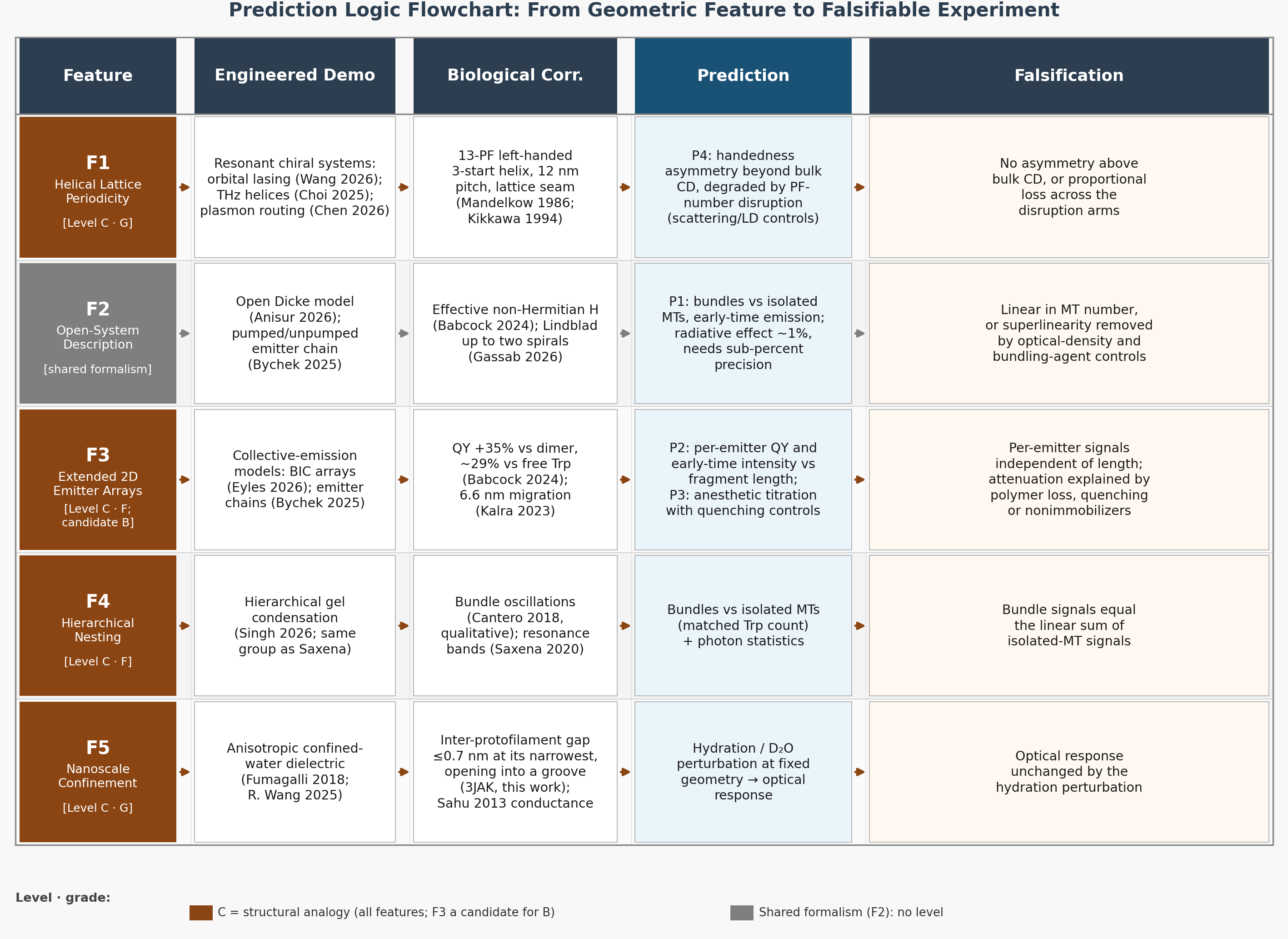
A methodological note applies across the optical predictions below. Open-system modeling of the tryptophan network shows that the direction and persistence of collective information flow depend strongly on the initial excitation condition: the preparation sets the balance between rapid bright (superradiant) export and slow dark (subradiant) retention, with a coherent, delocalized preparation retaining a subradiant component that survives the bright burst, whereas a maximally mixed preparation generates no sustained internal coherence (Gassab, Pusuluk, and Craddock, 2026). Experimental readouts should therefore fix and report the excitation/preparation condition, since the collective signature is initial-state-dependent; this makes the array-size and anesthetic measurements cleaner and forestalls ambiguity in interpretation. In vivo, the excitation source is itself unknown: the predictions above use external UV excitation, while the candidate endogenous source is ultraweak photon emission from reactive-oxygen chemistry, of which mitochondria are a major producer. Mould et al. (2023) reported that stressing isolated mitochondria altered respiration in a chemically separate, optically coupled population, but no photons were detected and the opaque barrier also blocks infrared and other radiation, so a light-mediated route remains a hypothesis.
Prediction 1: Inter-tubule coupling (Features 2 and 3). Hypothesis: If non-Hermitian coupling between adjacent emitter arrays drives collective emission in MT bundles, the bundle geometry creates an inter-tubule coupling that is absent in isolated MTs. This prediction is motivated by the prior observation (Cantero et al., 2018) that bundle assembly produces electrical oscillations richer than those the same group reported for 2D MT sheets; the present prediction asks whether an analogous bundle-level optical collective signature accompanies the previously reported bundle-level electrical signature. Open-system modeling adds a mechanistic rationale by analogy. Gassab, Pusuluk, and Craddock (2026) show that within a single microtubule spiral, neighboring tubulin units act as a structured reservoir that transiently stores and returns population and coherence (a non-Markovian backflow between subunits). That result is intra-microtubule, between tubulin units of one lattice rather than between separate microtubules; the bundle conjecture tested here is whether the same structured-reservoir coupling, scaled up to adjacent microtubules, produces a measurable inter-tubule optical signature. A reservoir that returns coherence to its subunits is the kind of environment in which bundling could matter. Experiment: Time-resolved UV fluorescence spectroscopy of (a) MT bundles of defined cross-section and (b) isolated MTs with matched total tryptophan count, measuring early-time emission intensity scaling and fluorescence decay kinetics, with an instrument response short enough to resolve any early-time component. Controls: Bundling changes more than inter-tubule coupling. It raises local optical density, scattering and reabsorption, alters the refractive-index environment, and the bundling agent (a crowding polymer, divalent cations or a bundling MAP) can itself change tryptophan emission. Samples should therefore be matched in optical density, with inner-filter and scattering corrections, absolute quantum yield (integrating sphere) and emission anisotropy measured alongside the decays, and the bundling agent applied to free tubulin and to isolated MTs below the bundling threshold. Expected outcome: Bundles should show superlinear early-time emission (superradiant burst signature) and altered decay timescales inconsistent with additive independent-emitter behavior, persisting after these controls. Falsification: Emission scales linearly with MT number and decay rates are unchanged by bundling, or any superlinearity disappears under the optical-density, scattering and bundling-agent controls. As for Prediction 2, a superlinear signal that survives the controls is necessary but not sufficient for an inter-tubule collective mode; it would still need agreement with a specified coupling model and, where feasible, a photon-statistics diagnostic. Feasibility: near-field coupling scales as r⁻³, so scaling the ~50–60 cm⁻¹ nearest-neighbor coupling at ~1.5 nm (Celardo et al., 2019; Babcock et al., 2024) gives about 3 cm⁻¹ for tryptophans 4 nm apart on neighboring MTs, about 0.2 cm⁻¹ at 10 nm and under 0.01 cm⁻¹ at 30 nm, far below the 50–300 cm⁻¹ disorder estimate (Table 2). A bundle effect would therefore have to come through shared radiative decay, which stays collective at these subwavelength separations; in the model of Babcock et al. (2024) it raises the thermal quantum yield by only about 1% between 10⁴ and 10⁵ tryptophans (a 91-microtubule bundle about 80 nm long), less than the reported quantum-yield uncertainties. The modeled yield had not saturated at 10⁵ tryptophans, and the yield calculation was not extended to bundles of micron-length microtubules (about 10⁶ tryptophans), so the estimate applies to short bundles. For longer bundles, the same study’s brightest-state enhancement, computed for bundles of 7 to 91 microtubules, saturates as they approach micron lengths (their Fig. 6). Dimensional scaling bounds what bundling could add beyond a single microtubule: in free space, an array’s largest collective decay rate keeps growing with emitter number only when the array extends beyond the wavelength in two or three dimensions, and then only as N1/4 or N1/3 (Mok et al., 2026). Treating a bundle as such an array is our approximation; on it, a bundle escapes single-microtubule saturation only once its cross-section approaches the wavelength, and even then the gain is sublinear. Prediction 1 therefore needs a time-resolved, per-emitter observable with sub-percent precision; otherwise it can bound, rather than detect, inter-tubule collective coupling.
Prediction 2: Array-size dependence (Feature 3). Hypothesis: If the 35% quantum yield enhancement over tubulin dimers and the anomalous exciton diffusion arise from collective behavior across the tryptophan array, rather than from reduced non-radiative decay upon assembly, then per-emitter observables (quantum yield, and early-time emission intensity divided by tryptophan count) should rise with lattice length up to an onset size and then saturate, whereas under additive independent emission they are independent of length; a purely classical reduced-quenching mechanism reaches its full effect as soon as a tryptophan is in its assembled local environment and predicts no such length dependence. A rising per-emitter dependence (superlinear total emission) is necessary for the collective account but not sufficient: as stated in Section 4.3, it would not by itself reject classical collective or optical-transport mechanisms, and discrimination would additionally require agreement with a specified collective dynamics model, exclusion of quantitatively constrained classical alternatives, and where feasible a coherence or photon-statistics diagnostic. The predicted direction is grounded on the biological side: Celardo et al. (2019) model a superradiant lowest-exciton state emerging above a ~12-spiral segment threshold via supertransfer between blocks, and the non-Hermitian eigenmode analysis of Gassab, Pusuluk, and Craddock (2026) finds that, in ordered lattices, the contrast between superradiant and subradiant lifetimes widens from a single dimer to 100 spirals. Experiment: Quantum yield and time-resolved UV fluorescence of MT fragments and short polymers of systematically varied length, compared against free tubulin and against the linear (additive) prediction. Because guanosine nucleotides confound tryptophan fluorescence (GTP absorbs in the 250–300 nm range, overlapping tryptophan excitation, and GTP-to-GDP hydrolysis alters tryptophan emission; Gassab and Craddock, 2026), nucleotide state must be held constant across the length series (for example, by using GMPCPP- or GDP-locked lattices) so that size-dependent scaling is not confounded by polymerization-coupled nucleotide changes. Expected outcome: Per-emitter quantum yield and early-time intensity rising with fragment length up to an onset size and then saturating, consistent with the onset modeled by Celardo et al. (2019) above ~12 spirals (~108 nm) of ordered lattice. Saturation is expected near the transition wavelength (λ ≈ 280 nm, about 31 spirals): in that model the largest superradiant decay width grows in proportion to the number of chromophores while the segment is no longer than the wavelength and begins to saturate beyond it, and in the model of Babcock et al. (2024) the quantum yield saturates once the microtubule reaches a few wavelengths. A general bound accounts for the saturation: in a one-dimensional array in free space much longer than the wavelength, the largest collective decay rate no longer grows with the number of emitters (Mok et al., 2026). The informative length series therefore runs from below ~100 nm to about 1 µm; in these models, longer polymers add no further per-emitter change. No engineered analog cited here supplies an onset size: in the BIC-metasurface model of Eyles et al. (2026), the second-order correlation is already about 1.5 for a 3 × 3 array (their Fig. 2a) and is set by the emitters’ coupling efficiency to the BIC mode (the β-factor) rather than by array size. Falsification: Per-emitter quantum yield, early-time intensity, decay components and anisotropy independent of fragment length across all sizes (total emission linear in tryptophan count). This would bound, not exclude, a collective contribution: dephasing can suppress the modeled onset, and bright and dark channels can redistribute decay without changing the integrated yield. Conversely, end effects, morphology-dependent scattering, reabsorption and multi-step transfer can produce a length dependence classically, so a positive result must be fitted jointly, across these observables, against independent-emitter, classical coupled-dipole or radiative-transfer, and open-quantum models, with the parameter ranges of Table 2 stated in advance.
Prediction 3: Anesthetic perturbation (Feature 3). Hypothesis: The tryptophan network geometry, not tryptophan chemistry alone, is the functionally relevant variable for the quantum yield enhancement reported by Babcock et al. Volatile anesthetics that bind to hydrophobic pockets adjacent to tryptophan residues (Craddock et al., 2017) should attenuate the QY enhancement (polymerized vs. free tubulin dimers), not the absolute QY alone. This prediction gains residue-level specificity from independent open-system modeling: Gassab, Pusuluk, and Craddock (2026) identify βW346 as the hub on which the network’s coherence and entanglement concentrate (it carries the dominant αW407–βW346 correlation channel when the network starts in either its superradiant or its most subradiant eigenstate), and the computed anesthetic binding site closest to a tryptophan (site 37, which persisted for the whole simulation) lies within 5.3 Å of βW346 (Craddock et al., 2012). The perturbation therefore targets the specific residue the correlation dynamics concentrate on, not merely a tryptophan near a binding pocket. Energy migration already responds: 50 μM etomidate and isoflurane shortened the migration length from 6.6 to 5.6 and 5.8 nm (Kalra et al., 2023). The prediction asks whether the quantum yield enhancement responds in the same way once the classical routes below are controlled. Computed halothane binding energies imply only 1–8% site occupancy at 1–2 MAC (Craddock et al., 2012), so an effect at clinical concentrations would have to arise from sparse binding. Experiment: Anesthetic titration, at clinically relevant and higher concentrations and across anesthetic types, into pre-assembled, stabilized MTs and matched free tubulin dimers, so that the anesthetic perturbs the lattice without changing how much of it forms; absolute QY is measured together with fluorescence lifetime and absorbance. Controls: Three classical routes could also lower the enhancement. Anesthetics can alter polymerization or MT stability, so polymer mass should be measured at each concentration (sedimentation or turbidity) and the enhancement normalized to it. Halothane quenches tryptophan fluorescence directly when it binds near the residue (Johansson et al., 1995; Solt et al., 2006), whereas Solt et al. (2006) describe sevoflurane, isoflurane and desflurane as anesthetics “which poorly quench tryptophan fluorescence”, although in their designed four-α-helix bundle protein these anesthetics still lowered tryptophan fluorescence through slower anesthetic-induced conformational changes. Quenching should therefore be measured on free tubulin dimers and on free tryptophan (or N-acetyl-tryptophanamide) and compared with the polymer, with lifetimes separating static from dynamic quenching. Anesthetic potency tracks lipophilicity, so nonimmobilizers of comparable lipophilicity (such as F6, 1,2-dichlorohexafluorocyclobutane; Koblin et al., 1994) should be included: if attenuation follows anesthetic potency rather than lipophilicity, they should have little effect, and the collective-mode calculation predicts a frequency shift of the opposite sign for F6 (Craddock et al., 2017). Expected outcome: Dose-dependent attenuation of the differential QY enhancement beyond what polymer loss and direct quenching account for, with potency ranking matching Craddock et al.’s hydrophobic binding affinity series and little effect from nonimmobilizers. Falsification: No correlation between anesthetic potency and differential QY enhancement; attenuation is non-selective; or the attenuation is fully accounted for by polymer loss or by quenching measured on monomer and free tryptophan, or is matched by nonimmobilizers. A potency-matched, quenching-corrected attenuation would show that anesthetic binding near βW346 acts on the assembly-dependent optical response (Grade F); it would not by itself identify a collective quantum mechanism, which would need the joint measurements set out in Section 4.3.
Prediction 4: Polarization and chirality disruption (Feature 1). Hypothesis: The MT helical lattice contributes a handedness-dependent asymmetry to UV fluorescence beyond the bulk circular dichroism of tubulin. The engineered chiral platforms of Section 4.1 are resonant and offer no mechanism at the microtubule’s wavelength-to-pitch ratio, so this is an open test of whether lattice chirality, entering through the coupling geometry of the helically arranged transition dipoles, is measurable at all. If it is, intact MTs should show such an asymmetry, and disrupting the lattice chirality (by altering protofilament number or seam structure) should degrade that asymmetry disproportionately to the magnitude of the structural change. The MT operates at λ/a ≈ 23 at the 280 nm absorption band and ≈ 28–29 at the 330–350 nm emission maximum, deep in the long-wavelength regime. Experiment: This prediction has two complementary arms. (i) Polarization signature: polarization-resolved fluorescence spectroscopy of intact MT assemblies vs. free (unpolymerized or depolymerized) tubulin dimers, with circular dichroism (CD) of the same samples as a bulk-chirality reference. (ii) Chirality disruption: a multi-arm lattice-perturbation design to control for the biochemical confounds of any single preparation — (a) GMPCPP-14PF vs GMPCPP-13PF MTs, varying PF number while holding GMPCPP biochemistry constant; (b) doublecortin-stabilized 13PF MTs vs unmodified mixed-PF preparations, varying PF distribution through MAP selection rather than nucleotide state; and (c) cross-species comparison using cell types with natively non-13-PF MTs (15-protofilament touch-receptor neuron MTs in C. elegans; Chalfie & Thomson, 1982) as in vivo controls. Controls: Chiroptical emission measurements on large assemblies carry known artifacts. Circular differential scattering by MTs and bundles can mimic a handedness asymmetry, so samples should be matched in turbidity and measured in a geometry that suppresses or corrects for scattering; linear dichroism, birefringence and photoselected linear polarization can leak into the circular channels, so linear anisotropy and linear dichroism should be measured on the same samples and the setup calibrated with a known circularly polarized emitter. Because the expected dissymmetry of tryptophan emission is small, the artifact floor must be established below the predicted effect. In arm (b), bound doublecortin adds its own aromatic emission, which must be measured on doublecortin alone and subtracted; in arm (c), tryptophan emission in cells is dominated by other proteins, so the comparison should use MTs isolated or reconstituted from those sources. Expected outcome: Intact MTs show asymmetric emission in left- vs. right-circular polarization channels exceeding the linear bulk-CD prediction; the asymmetry degrades disproportionately (a nonzero residual over the classical coupled-dipole prediction, defined below) in non-13-PF MTs across all three disruption arms. Falsification: No polarization asymmetry above the noise floor, or asymmetry fully accounted for by linear bulk-CD scaling; or polarization signatures scale proportionally with the structural difference across disruption arms (no geometric amplification); or natively non-13-PF MTs show polarization behavior indistinguishable from 13-PF MTs. Here ‘disproportionate’ is judged on absolute signed changes, not on a ratio of relative changes, which becomes unstable when either dissymmetry is near zero. For each disruption arm, the measured change in emission dissymmetry, Δg_lum, is compared with the change predicted for that measured structure (protofilament number, seam, bound MAPs) by a classical coupled-dipole calculation of the helically arranged tryptophan transition dipoles, which gives g_lum and g_CD together; the measured Δg_CD of the same samples checks that calculation. A fixed proportionality between g_lum and g_CD is not assumed, because nothing guarantees it across preparations that differ in nucleotide state, MAP binding, morphology and scattering. Uncertainties are propagated from replicate preparations, and the uncertainty and artifact models are specified before the measurement. The predicted outcome is a residual (measured minus classically predicted Δg_lum) whose confidence interval excludes zero and exceeds the artifact floor set by the controls; a residual whose interval includes zero, or that lies below that floor, is the falsifying case. Empirical constraint already in place: Kalra et al. (2023) tested GMPCPP-14PF against 13PF MTs in their exciton diffusion measurements and found similar diffusion lengths, indicating that GMPCPP biochemistry does not by itself abolish lattice-mediated phenomena; the polarization readout is a distinct observable, but this null result reduces the prior probability that observed GMPCPP-vs-taxol differences would be driven entirely by GMPCPP biochemistry. As with Predictions 1 and 2, a positive result would establish a geometry-dependent chiroptical effect (Grade F), not by itself a collective quantum one: coupled-oscillator models of chiral chromophore arrays predict lattice-dependent emission asymmetry classically, so the measured asymmetry must exceed what such a model gives for the same lattice before it counts toward a collective account.
5.4 Limitations
First, correspondence is not identity. The most important limitation is methodological: the feature-matching framework identifies geometric parallels but cannot establish that the same physical mechanism operates in both systems. A biological system with the right geometry could fail to exhibit collective quantum phenomena for reasons unrelated to geometry, such as ultrafast dephasing from protein dynamics, spectral inhomogeneity beyond the collective coupling threshold, or energetic disorder that localizes rather than delocalizes excitations.
Related to this: all the engineered chiral examples cited under Feature 1 are resonant, wavelength-scale structures, whereas the MT lattice sits deep in the long-wavelength regime (λ/a ≈ 23). Feature 1 is therefore a structural analogy without an engineered mechanistic anchor (Section 4.1); in particular, no direct analogy to the guided-resonance chiral lasing reported by Wang et al. (2026) is claimed.
A specific experimental-design caveat applies to the chirality-disruption arm of Prediction 4. The canonical 13-protofilament-to-14-protofilament comparison uses GMPCPP-stabilized MTs, which alters not only PF number but also nucleotide state and lattice spacing. Section 5.3 addresses this by specifying a multi-arm design (GMPCPP-PF-matched, MAP-stabilized, and cross-species comparisons) and notes that Kalra et al. (2023) have already shown GMPCPP-14PF and 13PF MTs to behave similarly in exciton diffusion measurements. Reviewers attentive to MT biochemistry are nevertheless correct that no single experiment fully decouples PF number from the surrounding biochemical variables, and the multi-arm design is a mitigation rather than a complete resolution.
Second, no direct quantum measurement has been made in intact MT assemblies. The biological evidence consists of bulk spectroscopic and electrical measurements (QY enhancement, energy migration distance, Raman line shape, bundle oscillation), together with computed anesthetic-induced mode shifts, whose interpretation in terms of collective quantum mechanisms is plausible but not uniquely established. The principal classical alternative, reduced non-radiative decay upon lattice assembly, is addressed at length in Section 4.3: current measurements do not exclude it, and an array-size scaling measurement (Prediction 2) is necessary but not sufficient to discriminate the accounts, because a superlinear dependence would not by itself reject classical collective or optical-transport mechanisms. Discrimination would also require a specified collective dynamics model, exclusion of quantitatively constrained classical alternatives, and where feasible a coherence or photon-statistics diagnostic. The remaining ambiguity is therefore one that the proposed experimental program is designed to narrow, not one that blocks interpretation.
Third, the five dimensions identified here are not exhaustive. Topological protection, defect tolerance, and phonon-bath engineering are geometric principles relevant to collective quantum phenomena in engineered systems that we have not systematically examined in the MT context. Our selection reflects the features most clearly supported by available biological measurements, not a complete survey of potentially relevant geometric properties. Because features were retained only when a microtubule counterpart existed (Section 2.1), their co-occurrence in microtubules is a property of the selection, not evidence. A reproducible scoping review with a defined search frame, screening record and negative comparators, and quantitative effect-size predictions for each model class in Section 5.3, are beyond this paper; both are natural next steps.
Fourth, three cited works are available only as preprints: two engineered studies (Eyles et al., 2026; Anisur & Choudhury, 2026) and the mitochondrial report of Section 5.5 (Yang et al., 2026). No correspondence level or conclusion depends on them; the candidate model correspondence for Feature 3 rests on peer-reviewed models (Section 4.3), although the BIC-mediated superradiance model closest in spirit to the MT tryptophan array awaits peer review.
Fifth, several of the engineered-system predictions invoked here rest on idealized, symmetric models, and the same caution applies to the microtubule side. Gassab and Craddock (2026) note that superradiance models built on symmetric tryptophan arrays from a single idealized structure (commonly PDB 1JFF) risk producing behavior that is an artifact of model symmetry rather than a robust biological feature, since residue orientations vary across species, nucleotide states, and conformations. The present framework is partly insulated from this concern because its claims rest on measured lattice-conditional behavior (quantum-yield enhancement, energy migration, bundle oscillations) and a computed anesthetic-potency correlation and on perturbation experiments performed on real, heterogeneous microtubules rather than on idealized-array predictions; the array-size and chirality-disruption experiments in Section 5.3 explicitly test native and variant lattices. Predictions that depend on idealized-array calculations should nonetheless be validated against structurally heterogeneous models before being treated as quantitative. Consistent with this caution, Gassab, Pusuluk, and Craddock (2026) find, in their non-Hermitian eigenmode analysis of large lattices, that static energetic disorder or MD-sampled structural disorder localizes and fragments superradiant coherence and compresses the bright–dark lifetime contrast, with superradiant states more sensitive than subradiant ones (an asymmetry established within this radiative-loss model, which does not include the non-radiative dephasing that operates in vivo), echoing Celardo et al.’s (2019) result that the ordered lattice tolerates disorder better than short-range geometries. The ordered models are nonetheless built on idealized 1JFF coordinates, so even these careful open-system treatments inherit the model-symmetry caveat, which is precisely why the discriminating experiments are specified on real, heterogeneous microtubules. A related scope limit applies to the whole modeling cluster invoked here (Babcock, Celardo, and Gassab–Pusuluk–Craddock): each treats radiative dissipation but omits the non-radiative dephasing that sets the functional coherence limit in a warm, aqueous environment. The lifetimes these models report are therefore radiative rather than functional, a further reason the decisive step is direct measurement rather than additional modeling.
Sixth, the local physical environment of the tryptophan network is not characterized at the scale that decides the question. The dielectric response near the chromophores, the static disorder in their site energies and how strongly it is correlated between neighbors, the bath spectral density, and the structure of wall-adjacent and luminal water are inferred from structures and simulations or assumed, not measured in intact microtubules (Table 2). The diagnostic grades in Section 4 are therefore ceilings set by current evidence: a feature graded F could still fail once these parameters are measured, and none can reach Q without measurements that constrain them. Pairing the predictions in Section 5.3 with direct measurements of these parameters, for example dielectric and hydration perturbations at fixed geometry, would narrow both uncertainties at once.
5.5 Other Biological Structures
Microtubules are not the only biological structures that exhibit some of these geometric features. Table 3 summarizes which features are structurally present in five candidate biological architectures and which combinations have been directly measured for collective behavior.
| Structure | F1 Helical periodicity | F3 Extended 2D emitter array | F4 Hierarchical nesting | F5 Nanoscale confinement | Direct measurement of collective behavior |
|---|---|---|---|---|---|
| Microtubule (13-PF) | ✓ | ✓ | ✓ | ✓ | partial¹ |
| DNA double helix | ✓ | ✓ | ✓ | ○ | partial² |
| Actin filament | ✓ | ○ | ✓ | — | — |
| Centriole | ✓ | (○)³ | ✓ | — | — |
| Vault particle | —⁴ | — | — | ✓⁴ | — |
| Icosahedral virus capsid | —⁵ | ✓⁶ | ○ | — | partial⁶ |
Legend: ✓ = structurally present and characterized; ○ = structurally present but functional role for collective behavior not established; — = not present or not applicable. In the last column, ✓ = collective behavior directly measured; partial = a related optical response measured without direct demonstration of a collective mode. The open-system description (Feature 2) is omitted because any of these structures admits one (Section 2.2).
Footnotes: (1) For microtubules, an assembly-dependent optical response has been measured (quantum yield, energy migration), but a collective mode has not; no direct measurement of collective quantum behavior in intact MT assemblies exists (Section 5.4). (2) Excitonic delocalization with a 1/e length of about three to four stacked bases has been measured in synthetic (dA)n oligonucleotides, with a larger fraction of more extended domains in A·T duplexes, while a substantial fraction of excited molecules decays as single bases do (Buchvarov et al., 2007); it has not been measured in the context of the full chromatin hierarchy. (3) Centrioles are typically built from nine triplet MTs arranged with ninefold symmetry, with doublets in Drosophila and singlets in C. elegans embryos (Winey & O’Toole, 2014); emitter-array properties are inherited from constituent MTs rather than emerging from centriole-level organization. (4) The vault shell has 39-fold dihedral symmetry (Tanaka et al., 2009) and narrows at its caps to 13-fold symmetry through a symmetry mismatch that leaves a narrow, greasy pore at each tip (Lövestam & Scheres, 2025); it is not a helical lattice, so Feature 1 is marked absent, and the pore is the confinement counterpart. (5) Virus capsids have icosahedral rather than helical symmetry. (6) Collective, superradiance-like emission has been reported in brome mosaic virus capsids densely labeled with a synthetic dye: under pulsed excitation, emission brightened nonlinearly with dye number and appeared as a delayed burst of about 20 ps (Tsvetkova et al., 2019; Holmes et al., 2022). A crowding agent (PEG) and a longer dye linker significantly disrupted the brightening, and dye on a silica bead of similar size showed none (Tsvetkova et al., 2019); photon statistics were not measured, and whether capsid symmetry itself is required remains open (Holmes et al., 2022). The emitter array is a synthetic dye rather than a native chromophore network. Actin filaments are helical polymers, with the main subunit contacts along a two-start helix (Holmes et al., 1990).
The icosahedral virus capsid is included as the one non-cytoskeletal entry because it shows that a protein template can support collective emission from an attached emitter array (Holmes et al., 2022). The role of the capsid’s icosahedral order has not been tested directly, so this case does not separate lattice regularity from helicity.
Mitochondrial cristae are a further candidate outside the cytoskeleton. A recent preprint, not yet peer reviewed, reports a 71.0 THz shoulder in the infrared spectra of living cells and isolated mitochondria that depends on intact mitochondrial structure, modelled as collective coupling between light and lipid CH₂ vibrations in the cristae membranes (Yang et al., 2026). The degrees of freedom differ from the microtubule case (mid-infrared lipid vibrations, not ultraviolet tryptophan excitons), so the observation bears on whether biological geometry shapes spectral response beyond isolated-molecule expectations, not on any microtubule mechanism.
What distinguishes microtubules here is less the co-occurrence of these features, which partly reflects how they were chosen (Section 2.1), than the availability of experimental measurements (Babcock, Kalra, Cantero, Sahu) and computational results (Craddock) that can be compared with engineered analogs. Whether other biological structures with subsets of these features also exhibit collective quantum signatures, and whether the number of simultaneously satisfied features correlates with the strength of collective behavior, is a question for future work. The geometric principles identified here may generalize beyond MTs; this paper is not the place to pursue that argument in full.
5.6 Consciousness
The relationship between microtubule quantum behavior and consciousness is the context in which most readers will have encountered this topic, and it deserves a direct statement. The present analysis neither requires nor evaluates consciousness-related interpretations. We do not claim that collective quantum phenomena in MTs contribute to consciousness, nor do we claim they do not. The question of whether MT quantum dynamics are causally relevant to neural information processing requires evidence that does not yet exist, specifically direct demonstration of collective quantum modes in intact neural microtubules under physiological conditions.
The geometric framework presented here is a step toward motivating the experiments that would generate such evidence. If the four predictions in Section 5.3 are tested and confirmed, the case for direct quantum measurement of MT collective modes would be substantially strengthened, and the relevance of those modes to broader functional questions could then be assessed on the basis of experimental data. We direct readers interested in the consciousness dimension to Hameroff and Penrose (1996) for the original theoretical proposal and to more recent critical discussions in the literature for the current state of that debate.
6. Conclusion
Microtubule architecture contains counterparts to four structural and boundary features of engineered nanophotonic systems reported or predicted to support collective quantum phenomena: helical lattice periodicity, extended two-dimensional emitter arrays, hierarchical nesting, and nanoscale confinement; models on both sides also share an open-system description, which is a common formalism rather than a correspondence. All four are structural analogies (Level C). For extended emitter arrays, the engineered and microtubule models share a collective-emission description, but its variables are not yet constrained on the microtubule side, and the measured assembly-dependent quantum yield and energy migration are consistent with, but do not demonstrate, a collective mode. The cited engineered chiral systems are resonant, far from the microtubule’s long-wavelength regime, and the nesting and confinement analogs share no quantitative model with the microtubule. On the diagnostic grades used here, the biological evidence for every feature is at most a classical-compatible functional response; none yet has a uniquely quantum diagnostic. Because the features were chosen for having microtubule counterparts, their co-occurrence is not offered as evidence; their value is that each identifies variables a discriminating experiment can perturb.
Comparative and predictive rather than conclusive, this analysis complements the dimensional convergence analysis of Echternach (2026), which identified a convergence of quantum-relevant phenomena across independent material systems on the 1–2 nm scale that MT geometry also occupies, and argued that this co-localization of scales motivates direct measurement. Together, the two analyses identify where to look and what to vary, not whether a collective quantum state exists: the candidate sites are the tryptophan-scale chromophore spacings, the hydrophobic pockets near them, and the structured interfacial-water layers adjacent to the protein wall, and the candidate mechanisms are those that the engineered analogs show can depend on such geometry. In those analogs geometry is an enabling condition, not a sufficient one; whether a collective mode forms in microtubules depends on the dielectric environment, disorder, bath coupling and excitation conditions (Section 2.4), which the geometry does not fix.
The decisive test is direct measurement. The four predictions in Section 5.3 (inter-tubule coupling signatures in MT bundles, superlinear array-size scaling of collective emission, anesthetic attenuation of the geometry-dependent quantum yield enhancement, and polarization/chirality-disruption signatures consistent with handedness-dependent emission) are screening experiments: Predictions 2–4 are within reach of existing spectroscopic methods, whereas Prediction 1 needs a sub-percent, time-resolved per-emitter observable and can otherwise only bound inter-tubule coupling. A null result bounds an assembly-dependent collective contribution within the sensitivity of the measurement and the parameter ranges of Table 2, rather than ruling it out; a positive result is compatible with classical coupled-dipole and transport accounts as well as a collective quantum one, so deciding between them additionally needs model-specific quantitative predictions and a quantum-diagnostic measurement. Until such measurements are made, the geometric framework presented here provides a more specific basis than prior treatments for designing those experiments and for interpreting their results within a physically grounded comparative context. That a 2026 review of cytoskeletal optics developed independently of this framework (Gassab and Craddock, 2026) leaves the same question open, whether the measured signatures need a quantum description, indicates that the discriminating experiments proposed here address a gap the field itself has identified rather than one peculiar to this analysis.
Data Availability Statement
All data analyzed in this study are derived from the published literature cited in the References section. Structural coordinates were obtained from the Protein Data Bank (PDB entries 1JFF and 3JAK). The analyses of these coordinates are two Jupyter notebooks that run offline on the deposited mmCIF files with NumPy, SciPy and gemmi: trp_distances.ipynb (tryptophan pair distances in 1JFF and 3JAK, Section 3.1; all 396 pairs and the input checksums) and 3JAK_cleft_figure.ipynb (inter-protofilament gap, Section 4.5 and Figure 2; its revised versions add the cutoff-sensitivity and narrowest-gap analysis and its sensitivity to grid spacing, slice width and end trimming). Both are provided to reviewers and will be deposited publicly with a DOI on acceptance.
Author Contributions
J.E.: Conceptualization, Methodology, Investigation, Writing – Original Draft, Writing – Review & Editing, Visualization.
Funding
This research received no external funding.
Acknowledgments
The author acknowledges the use of large language models as research assistants for literature organization, structural analysis, drafting, revision and pre-submission review, specifically Claude Opus 4.6, Claude Opus 4.7 and Claude Opus 5.5 (Anthropic) and ChatGPT 5.6 Sol (OpenAI), used in part through Synthesis Lab, a research-writing platform the author develops. All scientific claims, arguments, citations, and references were developed, verified, and validated independently by the author using Zotero reference management and direct analysis of deposited structural coordinates (PDB entries 1JFF and 3JAK). The structured five-feature architectural comparison between engineered nanophotonic systems and microtubule geometry represents original scholarly contribution.
Conflict of Interest
The author is the founder and developer of Synthesis Lab, a commercial research-writing platform, and has a financial interest in it. Synthesis Lab was used in preparing this manuscript (see Acknowledgments); it is not a subject of the study.
References
Anisur, S., & Choudhury, S. (2026). Dissipative Dicke time quasicrystals. arXiv preprint. https://doi.org/10.48550/arXiv.2602.05994
Babcock, N. S., Montes-Cabrera, G., Oberhofer, K. E., Chergui, M., Celardo, G. L., & Kurian, P. (2024). Ultraviolet superradiance from mega-networks of tryptophan in biological architectures. The Journal of Physical Chemistry B, 128(17), 4035–4046. https://doi.org/10.1021/acs.jpcb.3c07936
Bilezikian, D. L., Davidoff, D., Caram, J. R., Fay, T. P., Kalra, A. P., & Weiss, P. S. (2026). Wave mechanics of ciliary proteins: Quantum-enabled sensing and energy transfer in the “cellular antenna”. Aggregate, 7(6), e70354. https://doi.org/10.1002/agt2.70354
Buchvarov, I., Wang, Q., Raytchev, M., Trifonov, A., & Fiebig, T. (2007). Electronic energy delocalization and dissipation in single- and double-stranded DNA. Proceedings of the National Academy of Sciences, 104(12), 4794–4797. https://doi.org/10.1073/pnas.0606757104
Bychek, A., Holzinger, R., & Ritsch, H. (2025). Nanoscale mirrorless superradiant lasing. Physical Review Letters, 135(14), 143601. https://doi.org/10.1103/rbs2-2pd5
Cantero, M. del R., Villa Etchegoyen, C., Perez, P. L., Scarinci, N., & Cantiello, H. F. (2018). Bundles of brain microtubules generate electrical oscillations. Scientific Reports, 8(1), 11899. https://doi.org/10.1038/s41598-018-30453-2
Celardo, G. L., Angeli, M., Craddock, T. J. A., & Kurian, P. (2019). On the existence of superradiant excitonic states in microtubules. New Journal of Physics, 21(2), 023005. https://doi.org/10.1088/1367-2630/aaf839
Chaaban, S., & Brouhard, G. J. (2017). A microtubule bestiary: Structural diversity in tubulin polymers. Molecular Biology of the Cell, 28(22), 2924–2931. https://doi.org/10.1091/mbc.E16-05-0271
Chalfie, M., & Thomson, J. N. (1982). Structural and functional diversity in the neuronal microtubules of Caenorhabditis elegans. Journal of Cell Biology, 93(1), 15–23. https://doi.org/10.1083/jcb.93.1.15
Chen, Y., Chen, Y., Fang, Y., Ai, R., Cui, X., Zhuo, X., & Wang, J. (2026). Photonic spin-Hall effect in chiral plasmonic assemblies. Nature Communications, 17(1), 3246. https://doi.org/10.1038/s41467-026-70039-5
Choi, W., Moestopo, W. P., Gu, S., Lee, T., Yoo, J.-H., Xia, X., & Armstrong, M. R. (2025). Helical photonic metamaterials for encrypted chiral holograms. Advanced Science, 12(38), e07931. https://doi.org/10.1002/advs.202507931
Collini, E., Wong, C. Y., Wilk, K. E., Curmi, P. M. G., Brumer, P., & Scholes, G. D. (2010). Coherently wired light-harvesting in photosynthetic marine algae at ambient temperature. Nature, 463(7281), 644–647. https://doi.org/10.1038/nature08811
Craddock, T. J. A., St. George, M., Freedman, H., Barakat, K. H., Damaraju, S., Hameroff, S., & Tuszynski, J. A. (2012). Computational predictions of volatile anesthetic interactions with the microtubule cytoskeleton: Implications for side effects of general anesthesia. PLOS ONE, 7(6), e37251. https://doi.org/10.1371/journal.pone.0037251
Craddock, T. J. A., Kurian, P., Preto, J., Sahu, K., Hameroff, S. R., Klobukowski, M., & Tuszynski, J. A. (2017). Anesthetic alterations of collective terahertz oscillations in tubulin correlate with clinical potency: Implications for anesthetic action and post-operative cognitive dysfunction. Scientific Reports, 7(1), 9877. https://doi.org/10.1038/s41598-017-09992-7
Echternach, J. (2026). Dimensional convergence at the nanoscale: A novel synthesis of experimental evidence justifying direct measurement of quantum coherence in microtubules. Frontiers in Molecular Biosciences, 13, 1840116. https://doi.org/10.3389/fmolb.2026.1840116
Engel, G. S., Calhoun, T. R., Read, E. L., Ahn, T.-K., Mančal, T., Cheng, Y.-C., Blankenship, R. E., & Fleming, G. R. (2007). Evidence for wavelike energy transfer through quantum coherence in photosynthetic systems. Nature, 446, 782–786. https://doi.org/10.1038/nature05678
Eyles, D., Lassalle, E., Stokes, A., Nazir, A., & Paniagua-Domínguez, R. (2026). Dicke superradiance in extended 2D quantum arrays coupled to metasurface bound states in the continuum. arXiv preprint. https://doi.org/10.48550/arXiv.2602.04627
Fumagalli, L., Esfandiar, A., Fabregas, R., Hu, S., Ares, P., Janardanan, A., Yang, Q., Radha, B., Taniguchi, T., Watanabe, K., Gomila, G., Novoselov, K. S., & Geim, A. K. (2018). Anomalously low dielectric constant of confined water. Science, 360(6395), 1339–1342. https://doi.org/10.1126/science.aat4191
Gassab, L., & Craddock, T. J. A. (2026). Optical properties of the cytoskeleton: A review of light–matter interactions, quantum effects, and biological implications. Journal of Physics: Photonics, 8(1), 012006. https://doi.org/10.1088/2515-7647/ae3ee8
Gassab, L., Pusuluk, O., & Craddock, T. J. A. (2026). Quantum information flow in microtubule tryptophan networks. Entropy, 28(2), 204. https://doi.org/10.3390/e28020204
Guo, C., Wang, K., Zhang, L., Putzke, C., Chen, D., van Delft, M. R., Wiedmann, S., Balakirev, F. F., McDonald, R. D., Gutierrez-Amigo, M., Alkorta, M., Errea, I., Vergniory, M. G., Oka, T., Moessner, R., Fischer, M. H., Neupert, T., Felser, C., & Moll, P. J. W. (2025). Many-body interference in kagome crystals. Nature, 647(8088), 68–73. https://doi.org/10.1038/s41586-025-09659-8
Hameroff, S., Bandyopadhyay, A., & Lauretta, D. S. (2026). Microtubules are ‘fractal time crystals’: Implications for life and consciousness. Journal of Consciousness Studies, 33(1), 211–247. https://doi.org/10.53765/20512201.33.1.211
Hameroff, S. R., & Penrose, R. (1996). Orchestrated reduction of quantum coherence in brain microtubules: A model for consciousness. Mathematics and Computers in Simulation, 40(3–4), 453–480. https://doi.org/10.1016/0378-4754(96)80476-9
Holmes, K. C., Popp, D., Gebhard, W., & Kabsch, W. (1990). Atomic model of the actin filament. Nature, 347(6288), 44–49. https://doi.org/10.1038/347044a0
Holmes, J., Sushma, A. A., Tsvetkova, I. B., Schaich, W. L., Schaller, R. D., & Dragnea, B. (2022). Ultrafast collective excited-state dynamics of a virus-supported fluorophore antenna. The Journal of Physical Chemistry Letters, 13(14), 3237–3243. https://doi.org/10.1021/acs.jpclett.2c00262
Johansson, J. S., Eckenhoff, R. G., & Dutton, P. L. (1995). Binding of halothane to serum albumin demonstrated using tryptophan fluorescence. Anesthesiology, 83(2), 316–324. https://doi.org/10.1097/00000542-199508000-00012
Kalra, A. P., Benny, A., Travis, S. M., Zizzi, E. A., Morales-Sanchez, A., Oblinsky, D. G., Craddock, T. J. A., Hameroff, S. R., MacIver, M. B., Tuszyński, J. A., Petry, S., Penrose, R., & Scholes, G. D. (2023). Electronic energy migration in microtubules. ACS Central Science, 9(3), 352–361. https://doi.org/10.1021/acscentsci.2c01114
Kikkawa, M., Ishikawa, T., Nakata, T., Wakabayashi, T., & Hirokawa, N. (1994). Direct visualization of the microtubule lattice seam both in vitro and in vivo. Journal of Cell Biology, 127(6), 1965–1971. https://doi.org/10.1083/jcb.127.6.1965
Koblin, D. D., Chortkoff, B. S., Laster, M. J., Eger, E. I., II, Halsey, M. J., & Ionescu, P. (1994). Polyhalogenated and perfluorinated compounds that disobey the Meyer-Overton hypothesis. Anesthesia & Analgesia, 79(6), 1043–1048. https://doi.org/10.1213/00000539-199412000-00004
Lorenzoni, N., Lacroix, T., Lim, J., Tamascelli, D., Huelga, S. F., & Plenio, M. B. (2025). Full microscopic simulations uncover persistent quantum effects in primary photosynthesis. Science Advances, 11(40), eady6751. https://doi.org/10.1126/sciadv.ady6751
Lövestam, S., & Scheres, S. H. W. (2025). Cryo-EM structure of the vault from human brain reveals symmetry mismatch at its caps. Structure, 33(10), 1643–1648.e1. https://doi.org/10.1016/j.str.2025.07.014
Löwe, J., Li, H., Downing, K. H., & Nogales, E. (2001). Refined structure of αβ-tubulin at 3.5 Å resolution. Journal of Molecular Biology, 313(5), 1045–1057. https://doi.org/10.1006/jmbi.2001.5077
Mandelkow, E.-M., Schultheiss, R., Rapp, R., Müller, M., & Mandelkow, E. (1986). On the surface lattice of microtubules: Helix starts, protofilament number, seam, and handedness. Journal of Cell Biology, 102(3), 1067–1073. https://doi.org/10.1083/jcb.102.3.1067
Mok, W.-K., Poddar, A., Sierra, E., Rusconi, C. C., Preskill, J., & Asenjo-Garcia, A. (2026). Universal scaling laws for correlated decay of many-body quantum systems. Nature Physics, 22(10), 1691–1698. https://doi.org/10.1038/s41567-026-03448-4
Mould, R. R., Kalampouka, I., Thomas, E. L., Guy, G. W., Nunn, A. V. W., & Bell, J. D. (2023). Non-chemical signalling between mitochondria. Frontiers in Physiology, 14, 1268075. https://doi.org/10.3389/fphys.2023.1268075
Qin, Y., Chang, C.-W., Wang, L., & Zhong, D. (2012). Validation of response function construction and probing heterogeneous protein hydration by intrinsic tryptophan. The Journal of Physical Chemistry B, 116(45), 13320–13330. https://doi.org/10.1021/jp305118n
Reiter, G. F., Deb, A., Sakurai, Y., Itou, M., & Kolesnikov, A. I. (2016). Quantum coherence and temperature dependence of the anomalous state of nanoconfined water in carbon nanotubes. The Journal of Physical Chemistry Letters, 7(22), 4433–4437. https://doi.org/10.1021/acs.jpclett.6b02057
Rodríguez-Rubio, A., Zhu, H., Cheung, K. M., Rončević, I., Gödde, L. A., Hergenhahn, J., Field, J. L., Gupta, P., Stawski, W., Gotfredsen, H., Straw, J., Edmondson, M., O’Shea, J. N., Saywell, A., & Anderson, H. L. (2025). Synthesis of triple stranded porphyrin nanobelts. Science, 390(6770), 290–293. https://doi.org/10.1126/science.adz8900
Sahu, S., Ghosh, S., Ghosh, B., Aswani, K., Hirata, K., Fujita, D., & Bandyopadhyay, A. (2013). Atomic water channel controlling remarkable properties of a single brain microtubule: Correlating single protein to its supramolecular assembly. Biosensors and Bioelectronics, 47, 141–148. https://doi.org/10.1016/j.bios.2013.02.050
Saxena, K., Singh, P., Sahoo, P., Sahu, S., Ghosh, S., Ray, K., Fujita, D., & Bandyopadhyay, A. (2020). Fractal, scale free electromagnetic resonance of a single brain extracted microtubule nanowire, a single tubulin protein and a single neuron. Fractal and Fractional, 4(2), 11. https://doi.org/10.3390/fractalfract4020011
Shirmovsky, S. E., & Chizhov, A. V. (2023). Modeling of the entangled states transfer processes in microtubule tryptophan system. Biosystems, 232, 104967. https://doi.org/10.1016/j.biosystems.2023.104967
Singh, P., Sahoo, P., & Bandyopadhyay, A. (2026). Room-temperature soliton–polariton condensation in a hierarchical helical-nanowire fractal gel. Nanotechnology, 37(8), 085002. https://doi.org/10.1088/1361-6528/ae4755
Solt, K., Johansson, J. S., & Raines, D. E. (2006). Kinetics of anesthetic-induced conformational transitions in a four-α-helix bundle protein. Biochemistry, 45(5), 1435–1441. https://doi.org/10.1021/bi052206o
Tanaka, H., Kato, K., Yamashita, E., Sumizawa, T., Zhou, Y., Yao, M., Iwasaki, K., Yoshimura, M., & Tsukihara, T. (2009). The structure of rat liver vault at 3.5 angstrom resolution. Science, 323(5912), 384–388. https://doi.org/10.1126/science.1164975
Tsvetkova, I. B., Anil Sushma, A., Wang, J. C.-Y., Schaich, W. L., & Dragnea, B. (2019). Radiation brightening from virus-like particles. ACS Nano, 13(10), 11401–11408. https://doi.org/10.1021/acsnano.9b04786
Vivian, J. T., & Callis, P. R. (2001). Mechanisms of tryptophan fluorescence shifts in proteins. Biophysical Journal, 80(5), 2093–2109. https://doi.org/10.1016/S0006-3495(01)76183-8
Wang, M., Lv, N., Zhang, Z., Chen, Y., Si, J., Chen, J., Tang, C., Yin, X., Liu, Z., Xin, D., Yi, Z., Zheng, W., Kivshar, Y., & Peng, C. (2026). Chiral orbital lasing in a twisted bilayer metasurface. Nature Communications, 17(1), 2369. https://doi.org/10.1038/s41467-026-69665-w
Wang, R., Souilamas, M., Esfandiar, A., Fabregas, R., Benaglia, S., Nevison-Andrews, H., Yang, Q., Normansell, J., Ares, P., Ferrari, G., Principi, A., Geim, A. K., & Fumagalli, L. (2025). In-plane dielectric constant and conductivity of confined water. Nature, 646(8085), 606–610. https://doi.org/10.1038/s41586-025-09558-y
Winey, M., & O’Toole, E. (2014). Centriole structure. Philosophical Transactions of the Royal Society B: Biological Sciences, 369(1650), 20130457. https://doi.org/10.1098/rstb.2013.0457
Yang, Y., Gu, Z., & Song, B. (2026). A quantum state of mitochondria in the living cell. bioRxiv preprint. https://doi.org/10.64898/2026.09.10.750628
Zhang, R., Alushin, G. M., Brown, A., & Nogales, E. (2015). Mechanistic origin of microtubule dynamic instability and its modulation by EB proteins. Cell, 162(4), 849–859. https://doi.org/10.1016/j.cell.2015.07.012
Zhang, W., Craddock, T. J. A., Li, Y., Swartzlander, M., Alfano, R. R., & Shi, L. (2022). Fano resonance line shapes in the Raman spectra of tubulin and microtubules reveal quantum effects. Biophysical Reports, 2(1), 100043. https://doi.org/10.1016/j.bpr.2021.100043
Zhang, L., Wang, L., Kao, Y.-T., Qiu, W., Yang, Y., Okobiah, O., & Zhong, D. (2007). Mapping hydration dynamics around a protein surface. Proceedings of the National Academy of Sciences, 104(47), 18461–18466. https://doi.org/10.1073/pnas.0707647104